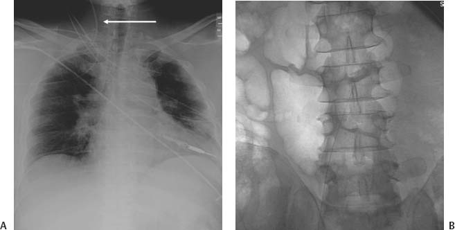
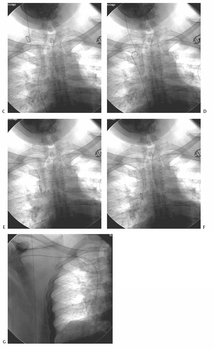
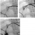
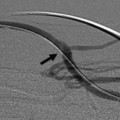
CASE 75 A 60-year-old male with a new diagnosis of lung cancer underwent surgical placement of a right subclavian chest port at an outside hospital. He then presented to our hospital for further treatment. A chest radiograph was obtained. Figure 75-1 Repositioning a malpositioned chest port catheter. (A) Chest radiograph shows catheter (arrow) misdirected into the right internal jugular vein. (B) Fluoroscopic image shows the guidewire advanced toward the heart from common femoral vein approach. (C) Fluoroscopic image shows the pigtail catheter adjacent to the port catheter tip in the right internal jugular vein. (D) Fluoroscopic image shows the pigtail catheter entangled with the port tip. (E–G) Fluoroscopic images show pigtail catheter redirecting the port tip into proper position. An upright chest radiograph showed a right subclavian chest port with the catheter portion of the device coursing cephalad into the right internal jugular vein. Chest port catheter malposition. The patient was brought to the interventional radiology suite, and the right groin was prepped. A 5-French (F) pigtail catheter was inserted into the right common femoral vein and advanced from the groin toward the heart (Fig. 75-1A). The loop of the catheter was advanced into the right internal jugular vein (Fig. 75-1B
Clinical Presentation
Radiologic Studies
Diagnosis
Treatment
![]()
Stay updated, free articles. Join our Telegram channel

Full access? Get Clinical Tree


