CASE 75 An adolescent girl presents with intermittent abdominal pain and a lower abdominal mass. Figure 75A Figure 75B Figure 75C Figure 75D On the axial T1-weighted image (Fig. 75A), two hyperintense structures, which represent a double vagina, are seen posterior to the bladder. On the axial T2-weighted image (Fig. 75B), the fluid collections in the vagina posterior to the bladder have varied signal, indicating subacute and more recent hemorrhage. (A second separate uterus not shown on these images was also present.) On the sagittal T1-weighted image (Fig. 75C), one vagina with very bright signal is seen to rise out of the pelvis. Another high-signal area seen posterior to the uterus, superior to the bright vaginal fluid in Fig. 75C, is a dilated fallopian tube. There is a small uterus anterosuperior to the vagina, which has similar signal to muscle on T1- and T2-weighted images (Fig. 75D). Hematocolpos, hematosalpinx, a double vagina with obstruction, and uterus didelphys
Clinical Presentation
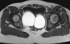
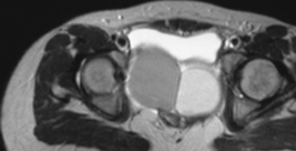
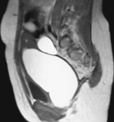
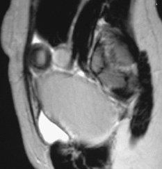
Radiologic Findings
Diagnosis
Differential Diagnosis
Discussion
Background
Related posts:


Stay updated, free articles. Join our Telegram channel

Full access? Get Clinical Tree


