Case 76
 Clinical Presentation
A 55-year-old man with long-standing scrotal swelling. There is a past history of scrotal trauma.
 Imaging Findings
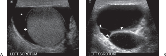
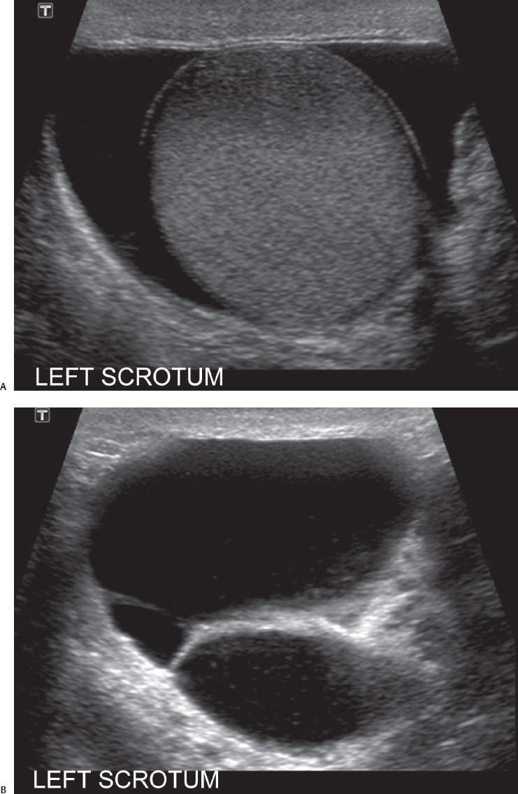
(A) Transverse sonographic image of the left scrotum shows a normal-appearing testicle (arrow) surrounded by clear fluid (asterisk) in the tunica vaginalis. (B) Transverse sonographic image of the left scrotum at a level below that in Figure A shows multiple thick septa (arrowheads) in the fluid. The fluid also shows some internal echoes.
Stay updated, free articles. Join our Telegram channel

Full access? Get Clinical Tree


