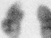
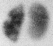
CASE 76 A 2-year-old boy presents having had a few episodes of urinary tract infection (UTI) 6 months earlier. Figure 76A (See Color Plate 76A.) Figure 76B (See Color Plate 76B.) Posterior (Fig. 76A) and left posterior oblique (Fig. 76B) images from a technetium Tc 99m dimercaptosuccinic acid (DMSA) study show a photopenic defect at the upper pole of the left kidney. A suspicious area is also seen laterally in the mid-pole of the left kidney proven to be a real defect on the left oblique images. As this boy had had no UTI for 6 months, these photopenic areas on DMSA represent renal scarring. The right kidney is normal. Renal cortical scarring on 99mTc-DMSA scan UTI is a common problem in pediatrics. It has been estimated that 8% of girls and 2% of boys will have a UTI during childhood. 99mTc-DMSA binds to the proximal convoluted tubules, resulting in fixation of the isotope and an unchanging image over many hours (hence the term static renal scan). Less than 10% of the injected dose is excreted in the urine. The delayed static images after intravenous injection represent functioning cortical mass and provide an accurate image of parenchymal outline. Posterior and oblique images are routinely obtained. Anterior views with the bladder empty are necessary when an ectopic or horseshoe kidney is suspected. Each image takes up to 5 minutes to acquire, and so sedation may be needed in a small number of children. DMSA studies are employed in some centers during an acute UTI to differentiate pyelonephritis from lower UTI. In many units, however, DMSA scans are reserved for assessing permanent renal scarring 3 to 6 months after a documented UTI.
Clinical Presentation
Radiologic Findings
Diagnosis
Differential Diagnosis
Discussion
Background
Etiology
Related posts:


Stay updated, free articles. Join our Telegram channel

Full access? Get Clinical Tree


