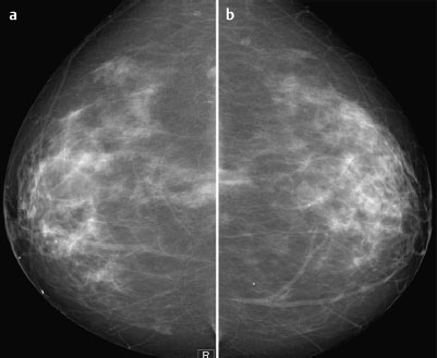
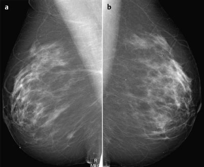
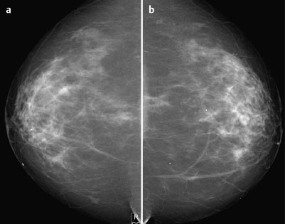
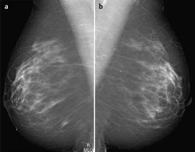
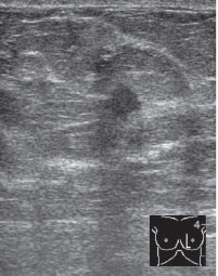
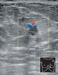
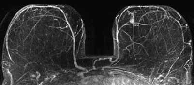
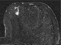
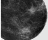
Case 77 (Continuation of Case75) Ah-ha, here are the earlier mammograms from Case 75, Fig. 77.1 a,b Digital mammography performed 1 year previously, CC view. Fig. 77.2a,b Digital mammography performed 1 year previously, MLO view. Fig. 77.3a,b Current mammography, CC view. Fig. 77.4a, b Current mammography, MLO view. Do the earlier mammograms change your interpretation of the recent findings? Between the inner quadrants of the left breast there was a microlobulated mass measuring 6 mm and disturbing a Cooper’s ligament (Fig. 77.5). This mass also showed increased peripheral vascularization (Fig. 77.6). Solitary hypervascularized lesion in the lower inner quadrant of the left breast (Fig. 77.7) with adjoining intraductal tumor component within a milk duct (Fig. 77.8). Spiculated, highly suspect lesion with internal calcifications (Fig. 77.9). Additionally, fibroadenoma with regressive changes. (Note: This lesion showed no enhancement in MRI.) US-guided core biopsy. Invasive ductal carcinoma, G3. Fig. 77.5 Sonography. Fig. 77.6 Color-coded Doppler sonography. Fig. 77.7 Contrast-enhanced MR mammography. Maximum intensity projection. Fig. 77.8 MR mammography subtraction image. Linear enhancement in tumor region indicates extensive intraductal component. Fig. 77.9 Spot compression, inner quadrants of left breast.
just arrived by bike courier!


Ultrasound
MR Mammography
Spot compression mammography, left breast (MLO)
Procedure
Histology of the specimen
BI-RADS Categorization | ||
Clinical Findings | right 1 | left 1 |
Ultrasound | right 1 | left 5 |
Mammography | right 1 | left 4 |
Histology
Invasive ductal carcinoma (9 mm)+extensive intraductal component.
IDC pTIb, pNO, G3.
Stay updated, free articles. Join our Telegram channel

Full access? Get Clinical Tree


