Case 77
 Clinical Presentation
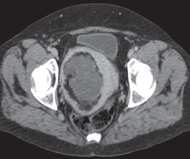
A 58-year-old woman with a pelvic mass.
 Imaging Findings
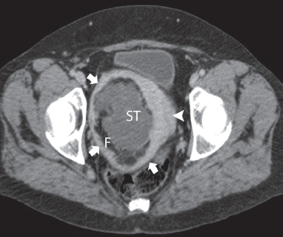
Contrast-enhanced computed tomography (CT) image shows a mass containing fat (F) and areas of soft-tissue (ST) attenuation within the pelvis. The mass is surrounded by a rim of soft tissue (arrows) that is continuous with the uterine myometrium (arrowhead) and has the same enhancement and attenuation.
 Differential Diagnosis
• Uterine lipoleiomyoma: A fat-containing mass within the uterus is characteristic. Myometrium surrounding the mass is confirmatory when present.
• Uterine lipoma:
Stay updated, free articles. Join our Telegram channel

Full access? Get Clinical Tree


