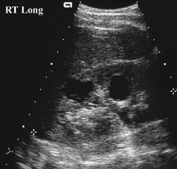
77 Autosomal Dominant Polycystic Kidney Disease

CASE 77


Clinical Presentation


A 6-year-old boy underwent abdominal ultrasonography because of a recent urinary tract infection (UTI). A positive family history of renal disease was present.


image

Figure 77A


Radiologic Findings


A longitudinal ultrasound image of the right kidney shows two anechoic lesions, both approximately 1.5 cm in diameter, with some posterior acoustic enhancement typical of renal cysts (Fig. 77A), which corresponded to the family history. The remaining right kidney and the left kidney were normal.


Diagnosis


Autosomal dominant polycystic kidney disease (ADPKD)


Differential Diagnosis


  • Multiple simple renal cysts (An occasional simple renal cyst may be seen in childhood, but multiple cysts do not normally occur, and the positive family history mentioned above is conclusive.)
  • Caliceal diverticula (Multiple diverticula would be very rare-one caliceal diverticulum may be indistinguishable from a simple renal cyst, but on IVU a caliceal diverticulum should opacify with contrast.)
  • Multicystic dysplastic kidney (is a congenital unilateral disorder, usually without normal parenchyma on the affected side)
  • Autosomal recessive polycystic kidney disease (manifests on ultrasound with bilateral large echogenic kidneys generally without resolvable cysts)
  • Tuberous sclerosis (TS)

Discussion


Background

Stay updated, free articles. Join our Telegram channel

Dec 21, 2015 | Posted by in PEDIATRIC IMAGING | Comments Off on 77 Autosomal Dominant Polycystic Kidney Disease

Full access? Get Clinical Tree

Get Clinical Tree app for offline access