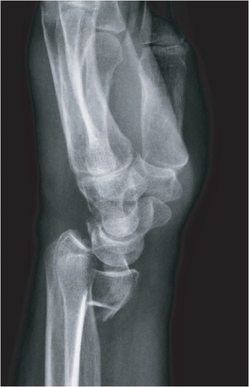
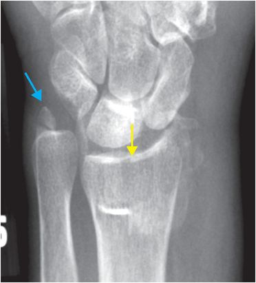
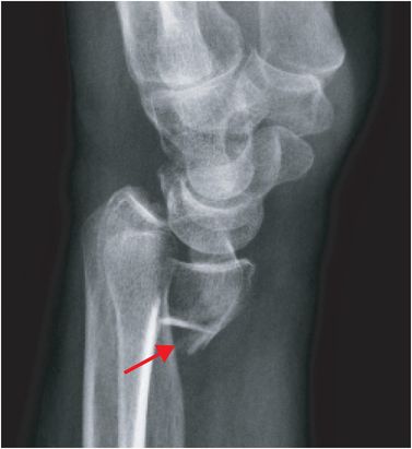
Diagnosis: Volar shear fracture of the distal radius (Volar Barton fracture)
Frontal and lateral radiographs of the wrist show a comminuted fracture of the distal radius with intra-articular extension to the radiocarpal articulation (yellow arrow) and volar displacement of the distal fragment (red arrow). There is also a fracture of the ulnar styloid (blue arrow).
Discussion
Overview of Barton fracture
Barton fracture is a shear-type fracture of the distal articular surface of the radius with translation of the distal radius fragment with the carpus. The volar and dorsal subtypes are distinguished by the direction of displacement of the radius fragment that articulates with the carpus.
Barton fractures are most commonly due to axial compression from a fall onto an outstretched hand (FOOSH), with the force vector transmitted through the distal radius. Affected patients are usually over 50 years of age.
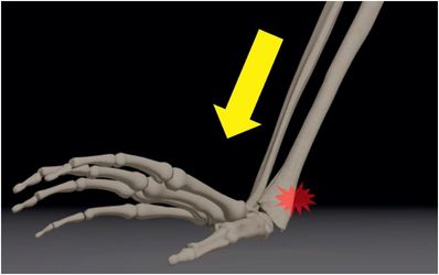
Axial loading at the wrist (arrow), commonly seen in a fall on an outstretched hand (FOOSH), causes the force vector to be transmitted to the distal radius with potential for distal radius fracture.
Classification of distal radius fractures
Multiple systems have been developed for classification of distal radius fractures. The Fernandez and Jupiter system (further discussed on the following pages) is the most commonly used classification among orthopedic surgeons since it addresses the mechanism of injury and accurately reflects prognosis.
Common named distal radius fractures
Several eponyms for distal radius fractures remain in common use, often described in conjunction with the Fernandez and Jupiter classification (indicated in parentheses).
Colles (type I) fracture is the most common wrist fracture of older adults, and is a bending-type fracture characterized by dorsal angulation of the distal fragment. Smith fracture is a reverse Colles, characterized by volar angulation of the distal fragment.
Barton (type II) fracture, as illustrated in the index case, is a shear-type intra-articular fracture of the distal radius. The carpus may be displaced in a volar or dorsal direction with respect to the distal radius.
Die-punch (type III) fracture is an impaction fracture of the lunate fossa of the distal radius.
Hutchinson (type IV) fracture, also known as chauffeur fracture, is an intra-articular radial styloid fracture. These are high-energy injuries often associated with radiocarpal dislocations and ligamentous injuries.
Imaging of distal radius fractures
PA and lateral wrist radiographs are most commonly obtained for initial evaluation, though additional external oblique view and PA view with the wrist in ulnar deviation may be necessary to evaluate the fracture morphology fully.
CT is helpful in characterizing fragment displacement and degree of articular surface involvement.
Treatment issues
Nonoperative management can be considered for minimally displaced or angulated Colles fractures.
Risk factors for instability include high degree of dorsal angulation, significant dorsal metaphyseal comminution, intra-articular extension, associated ulnar fracture, and age >60.
Closed reduction is attempted in all distal radial fractures, even those requiring immediate operative management. Operative fixation is recommended for Colles fractures with radial shortening >3 mm, dorsal angulation >10 degrees, or an intra-articular displacement or step off of >2 mm. It is therefore important to mention these findings if evident on radiographs.
It is critical for the clinician to evaluate for acute median nerve injuries or vascular compromise both pre- and post-reduction. Surgery is often required if neurovascular injury is suspected.
Related posts:
 12 68-year-old man with left lower quadrant pain and hypotension
 72 67-year-old female with shoulder pain and limited range of motion following a fall onto an outstretched hand
 35 37-year-old woman with a history of rheumatoid arthritis presenting with non-resolving bilateral effusions and chest pain
 62 38-year-old male complaining of diffuse abdominal pain after a motor vehicle collision
 53 42-year-old female presenting with fever and back pain
 66 21-year-old male with quadriplegia after diving into a shallow pond. The patient struck his head against an embankment, with his head flexed, chin against chest
Stay updated, free articles. Join our Telegram channel

Full access? Get Clinical Tree


