Case 79
 Clinical Presentation
A 53-year-old man with an indeterminate adrenal lesion on computed tomography.
 Imaging Findings
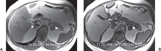
(A) In-phase axial T1-weighted magnetic resonance imaging (MRI) at the level of the adrenal glands shows that the left adrenal gland has been replaced by a nodule (arrow). The right adrenal gland is normal (arrowhead). (B) Opposed-phase axial T1-weighted MRI at the same level shows that the adrenal nodule has significantly and homogeneously lost its signal (arrow).
 Differential Diagnosis
• Adenoma of the adrenal gland: Uniform loss of signal on opposed-phase images is characteristic.
• Adrenal cyst: True adrenal cysts are rare. On T1-weighted images, their signal intensity may resemble that of an adenoma. However, on opposed-phase images, there is no loss of signal.
• Adrenal malignancy: Adrenal nodules may represent primary or secondary malignancy. However, malignant tissue is not expected to lose signal on opposed-phase images.
 Essential Facts
Stay updated, free articles. Join our Telegram channel

Full access? Get Clinical Tree


