CASE 8 A 51-year-old woman with a diagnosis of breast carcinoma and cervical carcinoma who is undergoing chemotherapy and radiation therapy for cervical carcinoma presents for staging (Figs. 8.1 and 8.2). Fig. 8.1 Fig. 8.2 • A 20 mCi dose of 99mTc-MDP is administered intravenously. • Whole-body images of the skeleton are obtained 3 hours after tracer administration. • A 1024 × 256 matrix is used for whole-body images. • Emphasize the importance of oral hydration to improve soft tissue and bladder clearance. No native kidneys are seen on anterior (Fig. 8.1) and posterior (Fig. 8.2) whole-body images. Normal soft tissue clearance is seen with appropriate osseous uptake. Within the right lower quadrant, focal uptake is shown overlying the right ilium. A degenerative/arthritic type of uptake is shown in the ankles, midfeet, and both first metatarsophalangeal joints. Mild uptake in the breast soft tissue is shown bilaterally.
Clinical Presentation
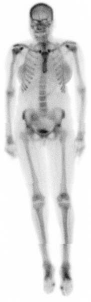
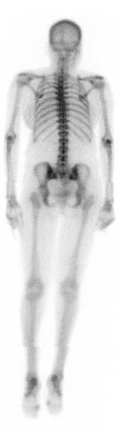
Technique
Image Interpretation
Related posts:


Stay updated, free articles. Join our Telegram channel

Full access? Get Clinical Tree


