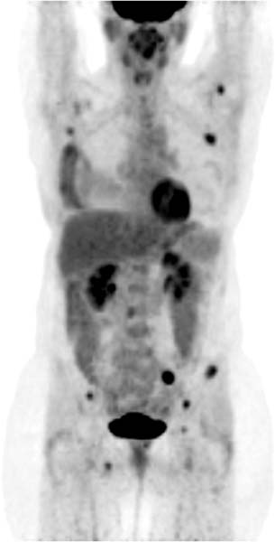
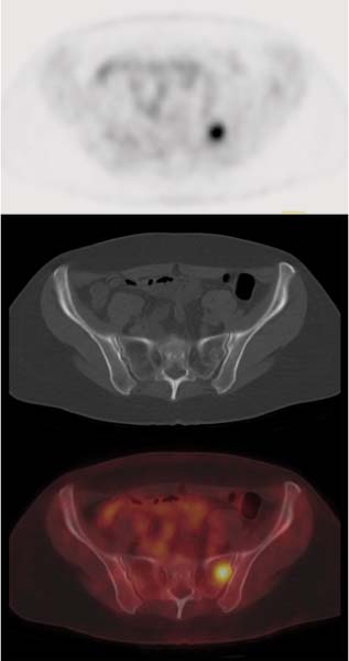
CASE 80 A 56-year-old woman presents with a palpable mass in the right breast and nipple inversion. Ultra-sound-guided biopsy reveals a poorly differentiated invasive ductal carcinoma. After radical mastectomy, 13 of 15 axillary lymph nodes are involved by tumor. PET/CT with 18F-FDG is performed postoperatively for further staging. Fig. 80.1 Fig. 80.2 • The patient is instructed to avoid vigorous exercise for 24 hours prior to the scan, fast for at least 4 hours before the study, drink water during the fasting and uptake periods, and void prior to the scan • A venous serum glucose sample is obtained prior to the injection of 18F-FDG (reference range is < 120 mg/dl for non-diabetic patients and < 200 mg/dl for diabetic patients), and the patient’s height and weight are recorded • Most PET scans are now acquired on a hybrid PET/CT scanner, and a low dose spiral CT is acquired for attenuation correction of the PET images and for anatomic localization. Some centers may choose to perform oral and/or intravenous contrast-enhanced diagnostic CT as part of the PET/CT examination • Sixty minutes following the 18
Clinical Presentation
Technique
![]()
Stay updated, free articles. Join our Telegram channel


Full access? Get Clinical Tree


