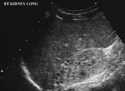
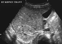
CASE 80 Initial abdominal sonogram in a male infant presenting with history of in utero oligohydramnios Figure 80A Figure 80B A highly echogenic kidney is evident on longitudinal (Fig. 80A) and transverse (Fig. 80B) ultrasound images with loss of the normal corticomedullary differentiation. Note the kidney is much more echogenic than the adjacent liver. A few small, peripheral, anechoic cysts are seen. Both kidneys have similar appearances and are small for age. Bilateral (cystic) renal dysplasia
Clinical Presentation
Radiologic Findings
Diagnosis
Differential Diagnosis
Discussion
Background
Related posts:


Stay updated, free articles. Join our Telegram channel

Full access? Get Clinical Tree


