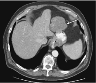
CASE 80 A 77-year-old man presents with abdominal pain. Fig. 80.1 Contrast-enhanced CT scan shows a lobulated, heterogeneously enhancing mass arising from the proximal stomach (arrow). An abdominal computed tomography (CT) scan (Fig. 80.1) shows a large, lobulated, heterogeneously enhancing mass arising from the proximal stomach. Gastrointestinal stromal tumor (GIST) GISTs are the most common mesenchymal tumors arising from the gastrointestinal (GI) tract wall. Nevertheless, they are uncommon, accounting for only 1 to 3% of all GI neoplasms, and usually affect patients > 50 years of age without any gender predilection. These tumors are also reported to occur in 10 to 25% of patients affected by neurofibromatosis 1. Previously mistaken for smooth muscle and neural tumors, GISTs show distinctive genetic and histologic features. They can occur anywhere from the rectum to the esophagus, the stomach being the most common site of origin; they can also be located in the mesentery, omentum, and retroperitoneum.
Clinical Presentation
Radiologic Findings
Diagnosis
Differential Diagnosis
Discussion
Background
Clinical Findings
Related posts:


Stay updated, free articles. Join our Telegram channel

Full access? Get Clinical Tree


