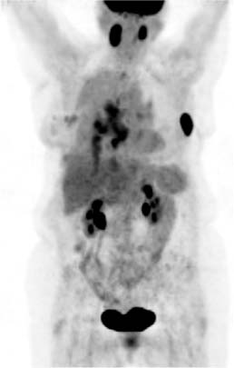
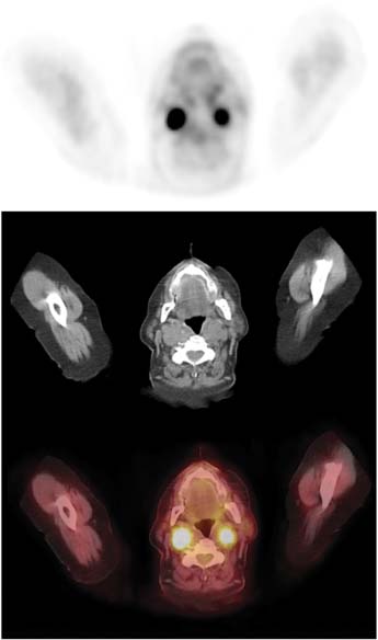
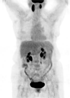
CASE 81 A 65-year-old woman has breast cancer. The patient was previously shown to have metastatic disease in the chest (Figs. 81.1 and 81.2). A re-staging examination after a course of chemotherapy was performed (Fig. 81.3). Fig. 81.1 Fig. 81.2 Fig. 81.3 • The patient is instructed to avoid vigorous exercise for 24 hours prior to the scan, fast for at least 4 hours before the study, drink water during the fasting and uptake periods, and void prior to the scan • A venous serum glucose sample is obtained prior to the injection of 18F-FDG (reference range is < 120 mg/dl for non-diabetic patients and < 200 mg/dl for diabetic patients), and the patient’s height and weight are recorded • A 10–20 mCi dose of 18F-FDG is injected intravenously (in a dimly lit quiet room for brain PET scans), and the patient is asked to rest comfortably in a room that is kept warm • Most PET scans are now acquired on a hybrid PET/CT scanner, and a low dose spiral CT is acquired for attenuation correction of the PET images and for anatomic localization. Some centers may choose to perform oral and/or intravenous contrast-enhanced diagnostic CT as part of the PET/CT examination • Sixty minutes following the 18
Clinical Presentation
Technique
![]()
Stay updated, free articles. Join our Telegram channel


Full access? Get Clinical Tree


