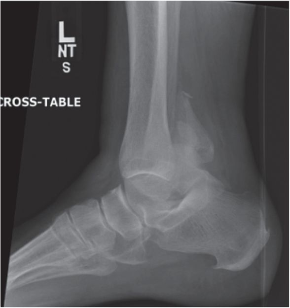
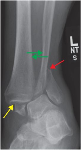
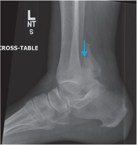
Diagnosis: Supination external rotation (SER) stage 4 ankle fracture
Frontal and lateral radiographs of the left ankle demonstrate displaced fractures of the medial (yellow arrow), lateral (red arrow), and posterior (blue arrow) malleoli, widening of the tibiofibular space (green arrows), and anterior and medial displacement of the tibia, consistent with SER stage 4 injury.
Discussion
The Lauge–Hansen classification system associates ankle injury types with mechanisms of injury, which allows consistent predictions of ligamentous injury patterns with each mechanism.
The classification system is based on two parts: the position of the ankle at the time of injury and the direction that the talus is displaced or rotated relative to the mortise.
The ankle may be in pronation (external rotation and eversion of the forefoot and abduction of the hindfoot) or in supination (internal rotation and inversion of the forefoot and adduction of the hindfoot) at the time of trauma. The three deforming forces are abduction, adduction, and external rotation. Therefore, the combined patterns of injury include supination adduction, supination external rotation (SER, as in the index case), pronation abduction, pronation external rotation, and pronation dorsiflexion.
The Danis–Weber classification is a much simpler classification of ankle fractures based on the location of the fibular fracture in relation to the tibiofibular syndesmosis.
Type A is an infrasyndesmotic fibular fracture, type B is a transsyndesmotic fibular fracture, and type C is a suprasyndesmotic fibular fracture. This simplified system is most helpful with type A and type C fractures, since in type A the syndesmotic ligaments are intact, and in type C the syndesmotic ligaments are disrupted. The status of the syndesmotic ligaments is indeterminate in type B fractures.
Approximately 40–70% of all ankle fractures are secondary to SER, which is the most common mechanism of ankle fracture. When the foot is in supination, the deltoid ligament is relaxed and there is tension on the lateral ligamentous structures; hence, the sequential and stepwise patterns of injury first affect the lateral side. Injury due to SER force is sequentially graded as follows:
SER stage 1: lateral rotation of the talus stresses and leads to the rupture of the anterior-inferior tibiofibular ligament (AITFL). It is considered a stable injury and is usually radiographically occult.
SER stage 2: increased stress on the talus leads to spiral fracture of the fibular malleolus, in the direction of low anterior to high posterior, at the tibiofibular syndesmosis. It is considered a stable injury. Patients with isolated fibular fractures at the level of syndesmosis (SER 2) require a gravity stress view for assessment of the deltoid ligament. If there is widening of the medial clear space (> 4 mm), deltoid ligament disruption should be suspected (deltoid ligament disruption upgrades the injury to SER stage 4).
SER stage 3: increased SER force causes injury to the posterior structures: rupture of the posterior-inferior tibiofibular ligament (PITFL) or fracture of the posterior malleolus of the tibia in addition to rupture of the AITFL and spiral fracture of the fibular malleolus (stage 1 and 2 injury).
SER stage 4: stage 4 injury is characterized by injury to the deltoid ligament / medial collateral ligament (MCL) complex or medial malleolus in addition to the lateral and posterior structures (stage 1–3 injuries). It is considered an unstable injury and requires open reduction more often than any other fracture type. If unrecognized, it can lead to talar subluxation, malunion, and arthrosis. If the diagnosis is suspected, follow-up radiographs or a stress series should be obtained to identify deltoid complex injury.
Clinical synopsis
The patient underwent open reduction and internal fixation using locking and nonlocking screws.
Related posts:
 12 68-year-old man with left lower quadrant pain and hypotension
 72 67-year-old female with shoulder pain and limited range of motion following a fall onto an outstretched hand
 35 37-year-old woman with a history of rheumatoid arthritis presenting with non-resolving bilateral effusions and chest pain
 62 38-year-old male complaining of diffuse abdominal pain after a motor vehicle collision
 53 42-year-old female presenting with fever and back pain
 66 21-year-old male with quadriplegia after diving into a shallow pond. The patient struck his head against an embankment, with his head flexed, chin against chest
Stay updated, free articles. Join our Telegram channel

Full access? Get Clinical Tree


