Case 83

 Clinical Presentation
A 17-year-old girl with a pelvic mass. She has not yet started menstruating.
 Imaging Findings
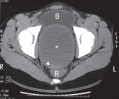
Contrast-enhanced axial computed tomography (CT) image obtained at the level of the pelvis. Between the urinary bladder (B) anteriorly and the rectum (R) posteriorly, there is a cystic abnormality (asterisk). It has a thick wall, and the contents are more hyperattenuating than the urine in the urinary bladder. There is higher-attenuation material posteriorly within the abnormality (arrowhead).
 Differential Diagnosis
• Hematocolpos: A large cystic abnormality within the pelvis in a young woman with delayed menarche should raise the suspicion of this abnormality. Because it represents distension of the vagina and uterus with blood, the location between the urinary bladder and rectum is characteristic. The contents consist of altered blood and have a higher attenuation than that of clear fluid. Areas of fresh blood with hematocrit effect may be seen, as in this case.
• Endometrioma:
Stay updated, free articles. Join our Telegram channel

Full access? Get Clinical Tree


