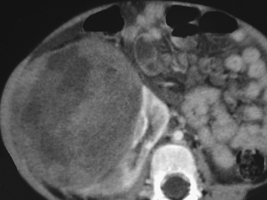
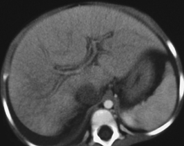
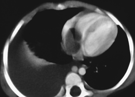
CASE 83 A 2-year-old girl presents with a palpable right-sided abdominal mass. Figure 83A Figure 83B Figure 83C Axial CT images after intravenous contrast enhancement show a large heterogeneous mass arising from the right kidney (Fig. 83A). More superior sections through the liver (Fig. 83B) and heart (Fig. 83C) show distended but non-opacified inferior vena cava (IVC), a right pleural effusion, and a filling defect in the right atrium due to tumor thrombus extension along the IVC into the right side of the heart. Note the enlarged azygos vein adjacent to the aorta on Fig. 83C. Wilms’ tumor with tumor thrombus in the IVC
Clinical Presentation
Radiologic Findings
Diagnosis
Differential Diagnosis
Discussion
Background
Related posts:


Stay updated, free articles. Join our Telegram channel

Full access? Get Clinical Tree


