CASE 84 A 3-year-old girl presents with a palpable hard lump in the buttock. Figure 84A Figure 84B Axial T1-weighted MRI before (Fig. 84A) and after (Fig. 84B) gadolinium enhancement shows a solid mass merging with the medial aspect of the left gluteus maximus muscle. This mass shows moderate heterogeneous enhancement after gadolinium administration. Rhabdomyosarcoma Rhabdomyosarcoma is the most common soft tissue sarcoma in children, representing 5 to 8% of all childhood cancers. Rhabdomyosarcoma is the most common malignant neoplasm of the pelvis in children, with a quarter of rhabdomyosarcoma cases affecting the genitourinary tract. These tumors can arise anywhere in the pelvis but in boys tend to occur predominantly in the bladder, prostate, and para-testicular sites (a so-called paratesticular location is applied to tumors arising in the spermatic cord, testis, epididymis, and penis). Paratesticular rhabdomyosarcoma accounts for 12% of scrotal tumors in childhood. The uterus and the vagina, in particular, are the more commonly affected organs in girls. Site of origin is critically important in determining prognosis in children with rhabdomyosarcoma. Tumors in the bladder/prostate area, for example, have a worse prognosis than the other genitourinary sites. Spread of disease is usually to regional lymph nodes initially, and it is important that these areas are scrutinized closely at all radiologic examinations. Routine staging at presentation with chest CT
Clinical Presentation
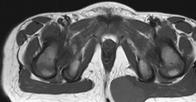
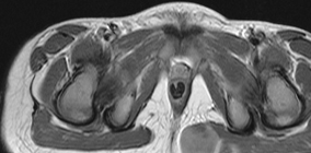
Radiologic Findings
Diagnosis
Differential Diagnosis
Discussion
Background
![]()
Stay updated, free articles. Join our Telegram channel


Full access? Get Clinical Tree


