Case 85

 Clinical Presentation
A 38-year-old man with bilateral flank masses. Computed tomography was performed.
 Imaging Findings
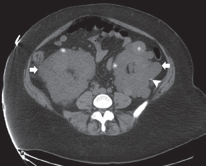
Noncontrast computed tomography image of the abdomen shows both kidneys (arrows) to be enlarged. The renal parenchyma on both sides has been replaced by multiple focal lesions with fluid attenuation. Scattered among these simple cystic focal lesions are other lesions with high attenuation (asterisks). Calcifications are seen in the wall of one of the cysts in the left kidney (arrowhead).
 Differential Diagnosis
• Adult polycystic kidney disease (PKD): Numerous, tightly packed renal cysts that involve both kidneys and replace the renal parenchyma are typical of adult PKD. Most of the cysts are typical simple cysts. However, they tend to hemorrhage, and the signal intensity may be variable depending on the amount and chronicity of hemorrhage. Calcifications in the walls of the cysts are an expected finding.
• Multiple bilateral simple cysts: Multiple simple cysts can occur in both kidneys and resemble adult PKD. They are fewer in number and sparser in distribution, and they do not displace the renal parenchyma. The kidneys are of normal size.
• Acquired PKD:
Stay updated, free articles. Join our Telegram channel

Full access? Get Clinical Tree


