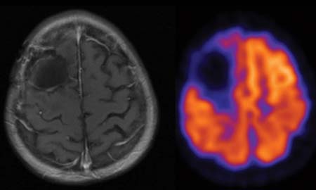
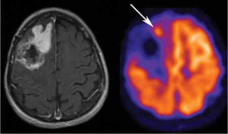
CASE 85 A 55-year-old woman undergoes resection of a glioblastoma multiforme from her right frontal lobe, followed by radiation therapy. Monitoring for tumor recurrence is performed with MRI and PET shortly (Fig. 85.1) and five months (Fig. 85.2) after treatment. Fig. 85.1 Fig. 85.2 Fig. 85.3 • The patient is instructed to avoid vigorous exercise for 24 hours prior to the scan, fast for at least 4 hours before the study, drink water during the fasting and uptake periods, and void prior to the scan • A venous serum glucose sample is obtained prior to the injection of 18F-FDG (reference range is < 120 mg/dl for non-diabetic patients and < 200 mg/dl for diabetic patients), and the patient’s height and weight are recorded • Most PET scans are now acquired on a hybrid PET/CT scanner, and a low dose spiral CT is acquired for attenuation correction of the PET images and for anatomic localization. Some centers may choose to perform oral and/or intravenous contrast-enhanced diagnostic CT as part of the PET/CT examination • Sixty minutes following the 18F-FDG injection, the spiral CT is performed using a weight-based algorithm from the skull base to the upper thighs (for most oncologic indications), or from the vertex to the feet, or centered over the brain depending on the tumor being evaluated • A 2D or 3D PET acquisition (depending on the manufacturer) is then performed over the same region (s), and all PET images are corrected for attenuation, detector efficiency, scatter, decay, and random coincidences • Consistency in 18F-FDG injected dose, scan acquisition time relative to the injection time, and acquisition and reconstruction protocols is important when performing serial scanning on the same patient • Gadolinium-enhanced MRI is also performed. T1-weighted post-contrast images are coregistered and fused with the PET data set. Axial MRI and corresponding PET image shortly after surgery and radiation therapy (Fig. 85.1) demonstrate absent 18F-FDG uptake in the site of surgical resection and decreased uptake in adjacent brain parenchyma secondary to radiation and edema. On follow-up MRI 5 months later (Fig. 85.2
Clinical Presentation
Technique
Image Interpretation
![]()
Stay updated, free articles. Join our Telegram channel


Full access? Get Clinical Tree


