Case 86
 Clinical Presentation
A 65-year-old man with an incidental renal finding on computed tomography of the chest.
 Imaging Findings
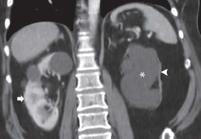
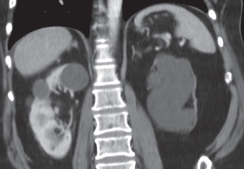
Coronal computed tomography (CT) image in the region of the kidneys shows a normal-size right kidney (arrow) with two simple cysts. The left kidney shows marked dilatation of the pelvis and calices (asterisk). There is nearly complete parenchymal atrophy (arrowhead). A dilated ureter is not visualized on this image and was not present on other images (not shown).
 Differential Diagnosis
• Left hydronephrosis due to ureteropelvic junction (UPJ) obstruction: The hydronephrosis is severe, and the renal parenchyma is markedly atrophic. The ureter is not dilated.
• Chronic left hydronephrosis due to ureteric obstruction:
Stay updated, free articles. Join our Telegram channel

Full access? Get Clinical Tree


