Case 88
 Clinical Presentation
A 54-year-old woman on long-term dialysis with the recent development of hematuria.
 Imaging Findings
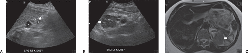
(A) Sonographic image of the right kidney demonstrates marked renal atrophy (arrow). Multiple simple-appearing cysts (asterisks) are seen in the kidney. (B) Sonographic image of the left kidney also demonstrates marked renal atrophy. Some cysts are seen. At the upper pole, there is a solid mass (arrow). (C) T2-weighted magnetic resonance imaging through the area of the mass confirms the presence of the mass (arrow) and shows it to be a solid lesion that is mildly hyperintense to muscle.
 Differential Diagnosis
Stay updated, free articles. Join our Telegram channel

Full access? Get Clinical Tree


