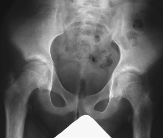
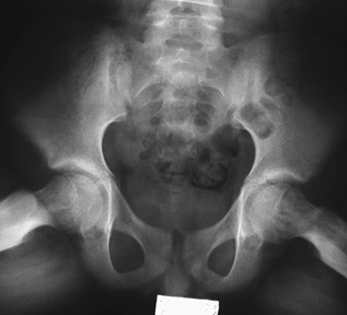
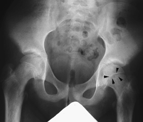
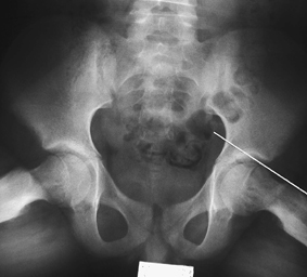
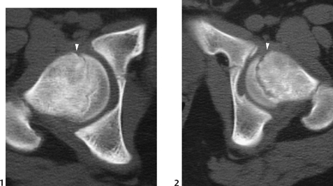
CASE 89 A 12-year-old obese boy presents with bilateral hip pain, worse on the left. Figure 89A Figure 89B The frontal film of the pelvis demonstrates widening of the femoral physeal plate on the left compared with the right (Fig. 89A). No portion of the left femoral epiphyseal center lies lateral to a line drawn tangential to the lateral border of the femoral neck. The “frog-leg” view demonstrates slight medial offsetting of the epiphyseal margins on underlying metaphyses (Fig. 89B), left greater than right. Figure 89C Frontal view of pelvis demonstrates relative equal size, configuration, and bone density of femoral head epiphyseal centers. The left physeal plate (arrowheads) is slightly more prominent than the right. Figure 89D A line drawn along the left lateral femoral neck does not intersect any portion of the femoral epiphyseal center, confirming the presence of “slip.” Slipped capital femoral epiphyses (SCFE) SCFE is the result of a fracture involving the proximal femoral physis. The apparent posterior and medial displacement of the epiphysis relative to the proximal metaphysis is the result of the lateral hip musculature pulling the femoral shaft anteriorly and laterally, giving a varus relationship. Rarely, a valgus slip is produced with superior and posterior displacement of epiphysis. SCFE is most often seen in adolescent males (2–4 male:female) who are often overweight. The age range is typically between 10 and 15 years, with the median age of males 2 years greater than females. SCFE occurs more commonly in African Americans than in whites and usually presents during a growth spurt. Bilateral epiphyseal slips are demonstrated 25% of the time. Approximately 60% of affected children have a skeletal maturity below the mean. Figure 89E (1
Clinical Presentation
Radiologic Findings
Diagnosis
Differential Diagnosis/Considerations
Discussion
Background
![]()
Stay updated, free articles. Join our Telegram channel


Full access? Get Clinical Tree


