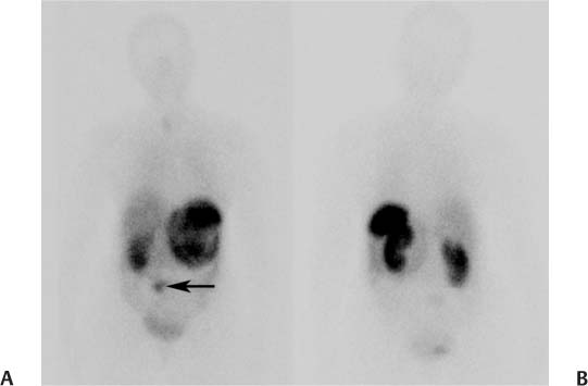
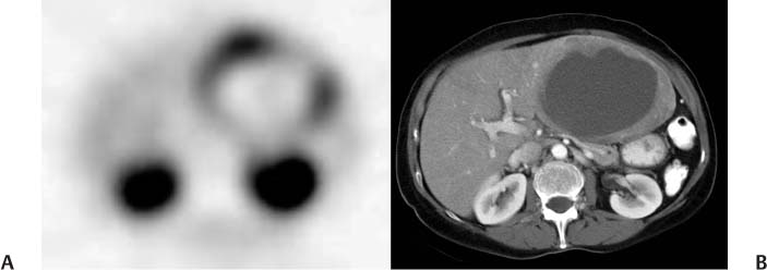
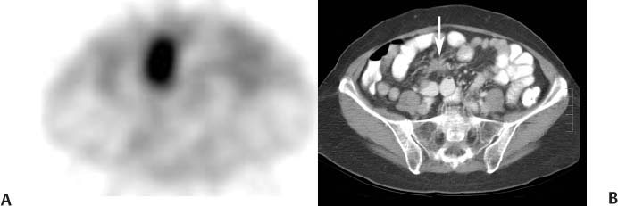
CASE 91 A 61-year-old woman presents with cramping and watery diarrhea that began 2 years earlier. More recently, she has experienced marked shortness of breath on exertion. CT of the chest, done for recurrent infections, reveals a large liver mass, biopsy of which leads to the investigations outlined below. She also undergoes echocardiography, which demonstrates marked thickening of the leaflets of the tricuspid and pulmonary valves with associated regurgitation, in addition to a dilated right ventricle with poor systolic function. Fig. 91.1 Fig. 91.2 Fig. 91.3 • 111In-pentetreotide • If possible, withhold nonradiolabeled octreotide before the scan: withhold 24 hours for short-acting octreotide and 3 to 4 weeks for long-acting formulations. • The patient should be well hydrated to enhance renal clearance. • If the patient has an insulinoma, a glucose infusion should be available to treat paradoxical hypoglycemia. • 6 mCi (222 MBq) • Slow intravenous injection over 1 minute • Medium-energy collimator • 172- and 247-keV photopeaks, 20% window • Planar: anterior and posterior views from head to pelvis at 4 and 24 hours • SPECT: abdomen and pelvis at 4 hours • SPECT: chest, abdomen, and pelvis at 24 hours • Additional images can be obtained at 48 hours if there is uncertainty whether abdominal activity represents pathologic or physiologic uptake. Anterior (Fig. 91.1A) and posterior (Fig. 91.1B) planar images at 24 hours demonstrate abnormal up-take in a large structure in the upper abdomen to the left of midline, and within a smaller structure lower in the abdomen (arrow) immediately to the right of midline. There is normal uptake in the spleen and kidneys (intense), liver and bladder (moderate), and thyroid (mild). An axial SPECT image at 24 hours (Fig. 91.2A) through the upper abdomen reveals marked pathologic uptake in the periphery of a large structure, confirmed on CT (Fig. 91.2B) to be a large metastasis with a necrotic center replacing most of the left lobe of the liver. There is normal intense physiologic activity in the kidneys. A SPECT image through the lower abdomen (Fig. 91.3A) demonstrates intense uptake in the primary tumor. This correlates with a spiculated mass (arrow) on CT (Fig. 91.3B) and an associated desmoplastic reaction in the mesentery. • Neuroendocrine tumor with a liver metastasis • Other malignancies expressing somatostatin receptors (eg, melanoma or breast carcinomas) • Inflammatory conditions expressing somatostatin receptors (eg, granulomatous diseases)
Clinical Presentation
Technique
Image Interpretation
Differential Diagnosis
Stay updated, free articles. Join our Telegram channel

Full access? Get Clinical Tree


