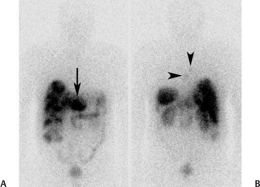
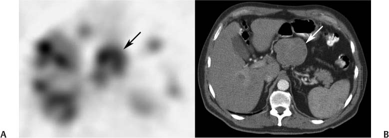
CASE 93 A 61-year-old man presents with recurrent duodenal ulcers and duodenitis. Fig. 93.1 Fig. 93.2 • 111In-pentetreotide • If possible, withhold nonradiolabeled octreotide before the scan: withhold 24 hours for short- acting octreotide and 3 to 4 weeks for long-acting formulations. • The patient should be well hydrated to enhance renal clearance. • If the patient has an insulinoma, a glucose infusion should be available to treat paradoxical hypoglycemia. • 6 mCi (222 MBq) • Slow intravenous injection over 1 minute • 172- and 247-keV photopeaks, 20% window • Planar: anterior and posterior views from head to pelvis at 4 and 24 hours • SPECT: abdomen and pelvis at 4 hours • SPECT: chest, abdomen, and pelvis at 24 hours • Additional images can be obtained at 48 hours if there is uncertainty whether abdominal activity represents pathologic or physiologic uptake. Anterior (Fig. 93.1A) and posterior (Fig. 93.1B) planar images at 24 hours demonstrate a large focus of intense uptake in the central abdomen, numerous foci of uptake throughout the liver, and foci of faint uptake in the thorax. A SPECT image (Fig. 93.2A) and corresponding CT slice (Fig. 93.2B) confirm uptake in a large mass arising from the head of the pancreas and within several liver lesions.
Clinical Presentation
Technique
Image Interpretation
Differential Diagnosis
Stay updated, free articles. Join our Telegram channel

Full access? Get Clinical Tree


