Case 94

 Clinical Presentation
A 38-year-old woman with severe lower abdominal pain.
 Imaging Findings

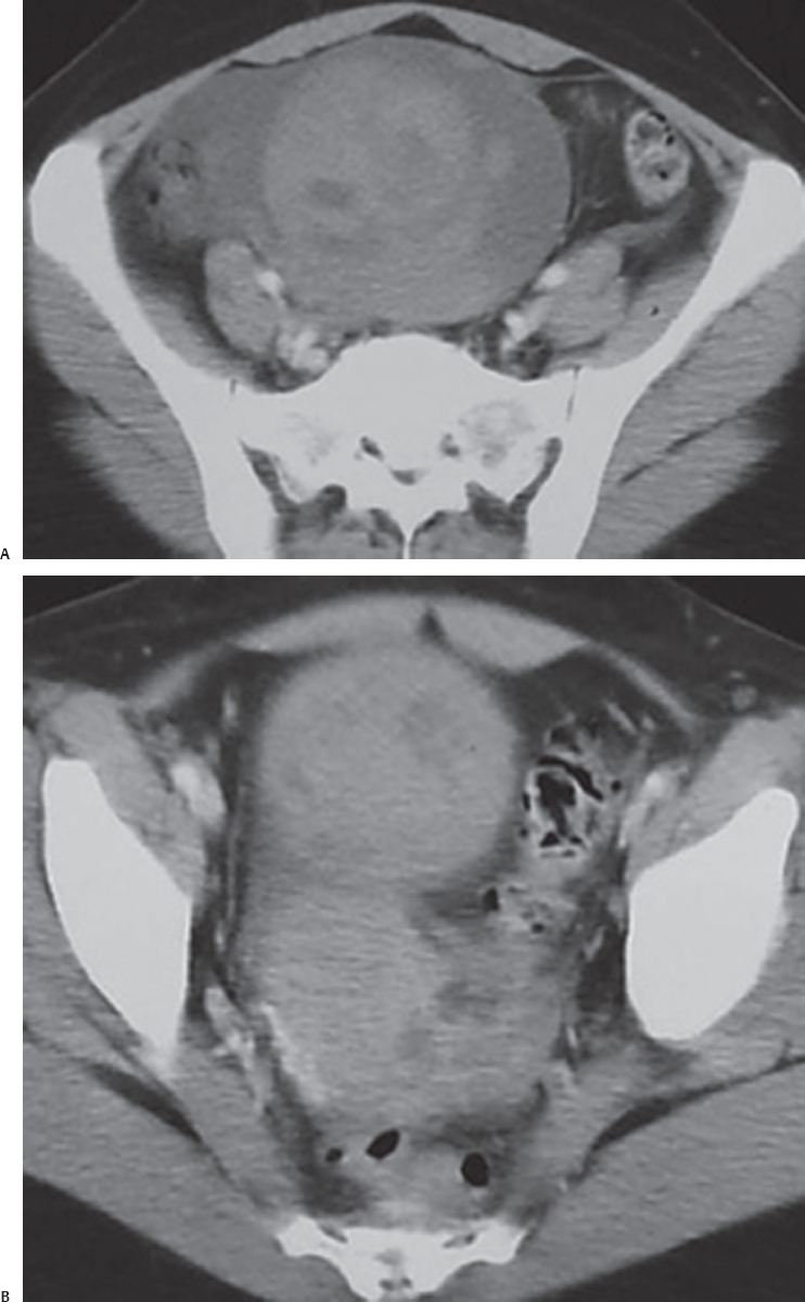
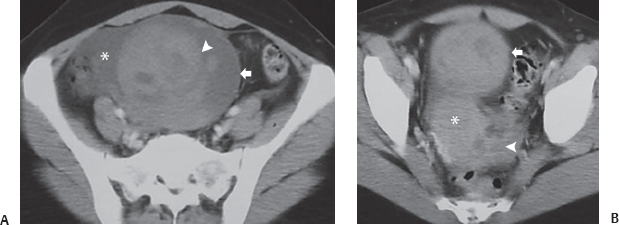
(A) Contrast-enhanced computed tomography (CT) of the pelvis shows a large round mass (arrow) in the midline of the pelvis, more toward the right. The mass has fluid attenuation, and swirling hyperattenuating areas (arrowhead) are seen within it. The wall of the mass is mildly thickened. The right ovary is not seen separately. There is free fluid in the right adnexal region (asterisk). (B) Contrast-enhanced CT of the pelvis at a level lower than that of Figure A shows the lower portion of the mass seen in Figure A (arrow). The uterus is seen behind the mass and is pulled toward the right side (asterisk). The left ovary appears normal (arrowhead).
 Differential Diagnosis


