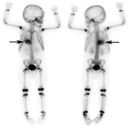
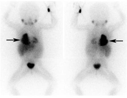
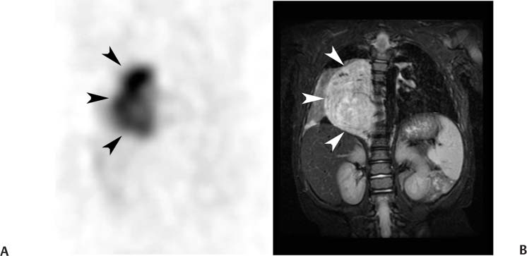
CASE 94 A 15-month-old girl has symptoms of an upper respiratory infection. A chest radiograph demonstrates a posterior mediastinal mass. She is referred for further evaluation of the mass. Fig. 94.1 Fig. 94.2 Fig. 94.3 Bone Scan (also see below for 123I-MIBG Scan) • 99mTc-HDP • No specific preparation • 6 mCi • Intravenous injection • Low-energy, high-resolution collimator • 140-keV photopeak, 20% window • Flow and pool images of chest, abdomen, and pelvis • Static images at 2 hours of entire skeleton 123I-MIBG • The patient should be well hydrated. • Drugs known or expected to interfere with MIBG uptake should be withheld. These include some β-blockers (in particular, labetalol), catecholamine agonists including oral decongestants, anti-psychotics, tricyclic antidepressants, some calcium channel blockers, and cocaine. A more detailed list, along with the recommended withholding period, is provided in Bombardieri et al. (2003). • Thyroid blockade should be performed with the administration of a saturated potassium iodide tablet, 65 mg once per day (pediatric dose), beginning on the day before the injection and continuing for a total of 3 days. • 1 mCi • Slow intravenous injection over 5 minutes • Low-energy, high-resolution collimator • 159-keV photopeak, 20% window • Static images at 18 hours of entire body • SPECT images of thorax and abdomen Anterior (Fig. 94.1A) and posterior (Fig. 94.1B) planar images from the bone scan demonstrate markedly increased uptake in the right mediastinal soft tissue mass, but no osseous abnormalities. Anterior (Fig. 94.2A) and posterior (Fig. 94.2B) planar images from the 123I-MIBG scan (Fig. 94.2) demonstrate intense accumulation of 123
Clinical Presentation
Technique
Technique
Image Interpretation
![]()
Stay updated, free articles. Join our Telegram channel

Full access? Get Clinical Tree


