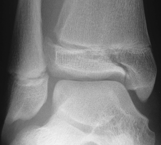
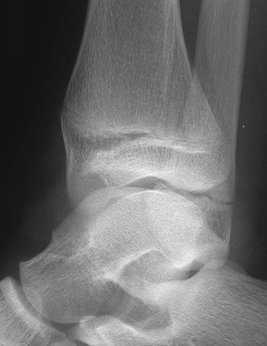
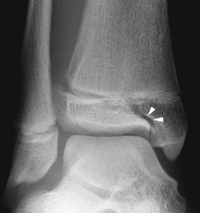
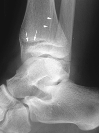
CASE 95 A 14-year-old athletic male presents with a swollen painful ankle after forced external version of foot. Figure 95A Figure 95B The frontal oblique radiograph shows a faint vertical lucent line, which extends through the distal tibial epiphysis (Fig. 95A). The lateral view shows an oblique fracture line extending through the distal tibial metaphysis posteriorly (Fig. 95B). Widening of the anterior and lateral physeal plate is also seen. Figure 95C Frontal oblique radiograph of the ankle demonstrates a lucent cleft (arrowheads) spanning the distal tibial epiphysis. There is widening of the lateral physeal plate. Figure 95D Lateral radiograph of ankle depicts widening of anterior physeal plate (arrows). A fracture line spans the distal tibial metaphysis posteriorly (arrowheads) with mild dorsal displacement of the metaphyseal fragment. Triplane fracture Tillaux fracture (no metaphyseal component) The distal tibial epiphysis is the second most common site for fractures involving the physeal plate with an estimated incidence of 60 in 10,000. The degree of skeletal maturity at the time of trauma plays the major role in the resulting fracture/injury pattern. Marmor in 1970 first described the complex nature and coronal, sagittal, and axial fracture components of the triplane fracture, which encompasses ~7% of ankle fractures in girls and ~15% in boys. There are several anatomic variants for triplane fractures consisting of two to four principle fragments. Triplane fractures occur in children between 11 and 16 years of age with a mean age in boys of 14.8 years and in girls 12.8 years. The triplane fracture and its variants are primarily the result of excessive external rotational forces placed on the ankle/foot. A degree of forced plantar flexion can also contribute to the pattern.
Clinical Presentation
Radiologic Findings
Diagnosis
Differential Diagnosis
Discussion
Background
Etiology
Pathogenesis
Related posts:


Stay updated, free articles. Join our Telegram channel

Full access? Get Clinical Tree


