Case 96
 Clinical Presentation
A 36-year-old man with long-standing subcutaneous and skin lesions presents with increasing abdominal girth.
 Imaging Findings
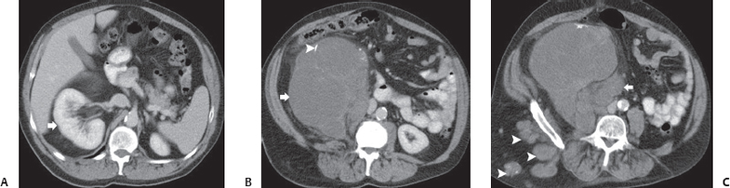
(A) Contrast-enhanced computed tomography (CT) image at the level of the kidneys shows the axis of the right kidney (arrow) to be altered from the normal vertical to a horizontal orientation. (B) Contrast-enhanced CT image at a level below that of Figure A shows that a large mass (arrow) in the right retroperitoneum is responsible for the change in the renal axis. It has solid and cystic areas and shows some calcifications (arrowhead). (C) Contrast-enhanced CT image at a level below that of Figure B shows more nodules and masses (arrow) in the retroperitoneum adjacent to the mass seen in the images above. In addition, nodules are seen in the subcutaneous tissue (arrowheads).
 Differential Diagnosis
• Plexiform neurofibromatosis of the retroperitoneum:
Stay updated, free articles. Join our Telegram channel

Full access? Get Clinical Tree


