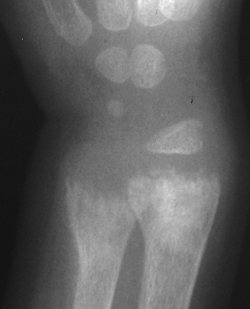
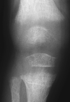
CASE 96 A short, lethargic 21/2-year-old boy presents with enlarged wrists and knees. Figure 96A Figure 96B A frontal view of the wrist (Fig. 96A) and knee (Fig. 96B) depicts widened physeal plates with cupping and fraying of subphyseal metaphysis. Rickets Rickets can be described as an alteration in the orderly mineralization and development of the growing skeleton generally related to defective vitamin D metabolism. The disease process was first recognized in the 1600s during the industrial revolution. Lack of sun exposure due to the smog, high buildings, and crowded streets in urbanized centers was the primary initiating factor at that time. In 1900 it was estimated that ~80% of children <2 years of age had evidence of rickets in the Boston area. The availability of synthesized vitamin D in the 1920s nearly eradicated vitamin D-deficiency rickets. The most common causes of rickets in the United States today are vitamin D-resistant or vitamin D-dependent disorders and renal osteodystrophy. In regions of Asia and Africa, the prevalence of vitamin D-dependent rickets is ~40%. There are >50 disease processes, with a vast spectrum of etiologies and clinical presentations, that can result in rickets. The causes of rickets can be categorized into the following groups:
Clinical Presentation


Radiologic Findings
Diagnosis
Differential Diagnosis
Discussion
Background
Etiology
Histology/Pathology
Stay updated, free articles. Join our Telegram channel

Full access? Get Clinical Tree


