A Brief History of Image-Guided Therapy


A Brief History of Image-Guided Therapy


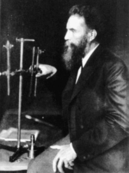
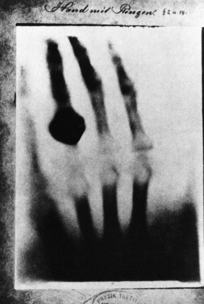
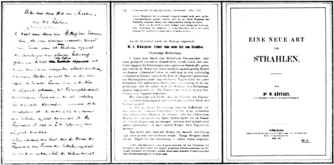
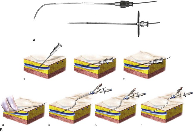
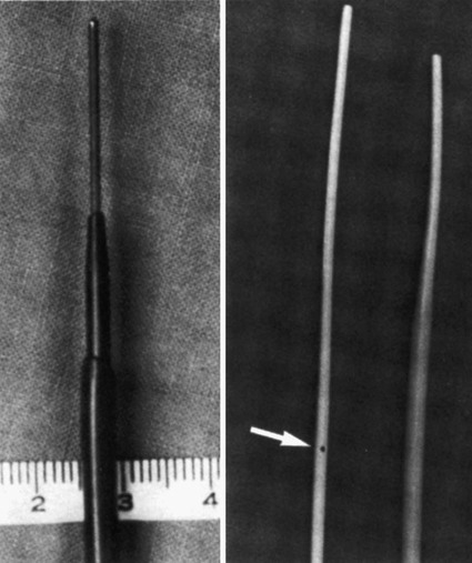
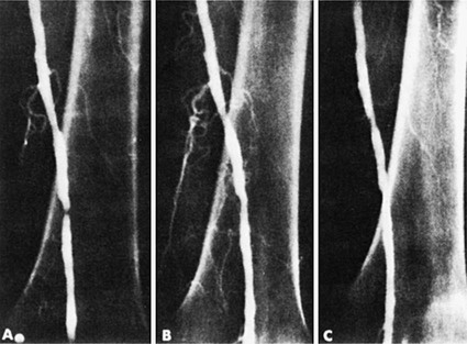
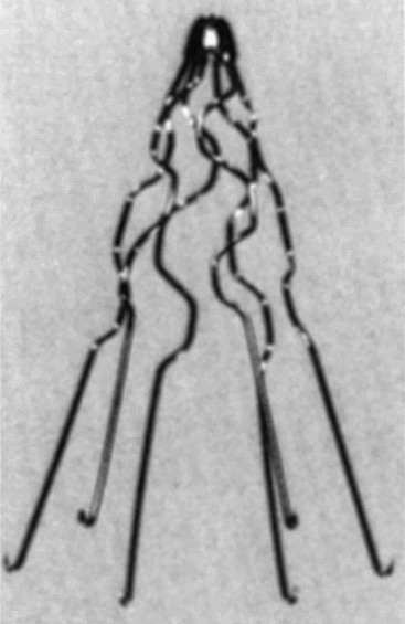
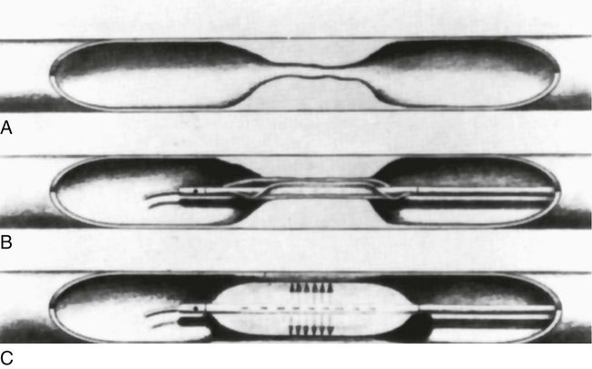
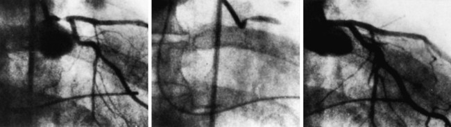
Anthony C. Venbrux, Jozef M. Brozyna, Suma Chandra, Hank K. Chen, Gina D. Tran and Dmitri A. Gagarin


Historical Highlights of Endovascular Therapy


• British dentist Charles Stent develops a plastic material for taking mouth impressions (i.e., creates a “scaffold”) (1856).


• Image guidance was made possible by the discovery of x-rays by Wilhelm Conrad Röntgen, November 8, 1895 (Figs. 1-1 to 1-3).


• Sven Ivar Seldinger develops percutaneous vascular catheterization (1952) (Figs. 1-4 and 1-5).


• Percutaneous revascularization is accomplished by Charles Dotter with coaxial dilators (1964) (Figs. 1-6 and 1-7).


• Transcatheter embolotherapy is performed by Charles Dotter to control acute upper gastrointestinal bleeding (1970).


• Lazar Greenfield pioneers caval interruption with a vena cava filter (1973) (Fig. 1-8).


• Andreas Gruentzig performs percutaneous angioplasty (1977) (Figs. 1-9 and 1-10).


• Chemoembolization was performed by Cato (1981).


• Julio Palmaz develops the endovascular balloon-expandable stent (1985).


• Juan C. Parodi, Julio C. Palmaz, and H. D. Barone develop stent-grafts (1990).


Endovascular Milestones


November 18, 1895, was a day of historical significance. In a physics laboratory in the southern part of Germany, Conrad Wilhelm von Roentgen accidentally discovered x-rays. The mysterious rays illuminated medical science, and radiology was born. From this serendipitous discovery, image-guided minimally invasive procedures evolved to provide patients with therapeutic options in the management of vascular and nonvascular diseases.


This chapter is neither comprehensive nor complete. Of the many potential technologic advances, arterial and venous endovascular therapy is the focus in this section. Later in the chapter, the history of nonvascular interventions is covered.


Arterial Endovascular Therapy: Revascularization and Vessel Reconstruction


An English dentist, Dr. Charles Stent, developed a thermoplastic material for taking impressions of toothless mouths in 1856. Thus the “stent” may be considered a “scaffold.” For purposes of this discussion, a stent is used for reconstruction in the vascular (or nonvascular) systems.


Percutaneous vascular catheterization as a viable endovascular technique was described in June 1952, when Sven Ivar Seldinger presented his idea of replacing an arteriography needle with a catheter. Translumbar aortography and direct carotid puncture were effectively relegated to historical descriptions in textbooks.


The technique of percutaneous revascularization advanced rapidly in 1964, when Charles Dotter used coaxial “pencil-point” dilators to treat a superficial femoral artery stenosis. The era of image-guided revascularization of the lower extremities had begun.


Although Dotter was successful in dilating femoral arterial stenoses, the use of coaxial “pencil-point” dilators (Van Andel Catheters [Cook Inc., Bloomington, Ind.]) required progressive enlargement of the percutaneous puncture site. Development of the angioplasty balloon by Andreas Gruentzig in 1977 resulted in another major step forward that allowed the percutaneous arterial entry site to be kept to a minimum (see Figs. 1-9 and 1-10). Early balloon designs were hampered by uneven balloon dilation and frequent rupture. Such events often led to pseudoaneurysm formation or dissections and resulted in vessel thrombosis.


The arterial metallic stent was invented in the 1980s by Julio Palmaz at the University of Texas Health Science Center in San Antonio. Dr. Palmaz described his stent in 1985 and continued work on his device in 1986. Later developments included the use of a stent design consisting of a single stainless steel tube with parallel staggered slots in the wall. When the stainless steel tube was expanded, the slots formed diamond-shaped spaces that resisted arterial compression. This design became the first stent approved by the U.S. Food and Drug Administration (FDA) for vascular use.


Stanley Baum and Moreye Nusbaum pioneered catheter embolization in the mid-1960s for the purpose of treating acute gastrointestinal bleeding. In 1970, Charles Dotter reported utilizing an autologous clot as the embolic agent to control acute upper gastrointestinal bleeding by selective embolization of the right gastroepiploic artery in a patient who was a poor surgical candidate. Robert White used this technique in 1974 to control bleeding duodenal ulcers when the hemorrhage was unresponsive to intraarterial injections of vasopressin.


Chemoembolization was largely pioneered by the Japanese urologist Cato in 1981. Cato used particles about 200 µm in size to demonstrate that chemoembolization with microcapsules containing chemotherapeutic agents was superior to local intraarterial injection of antitumor agents.1


Stent-grafts, pioneered by Juan Parodi and Charles Dotter, became the major impetus for future endovascular reconstruction procedures used to exclude an aneurysm, close an arteriovenous fistula, and reconstruct the central lumen of a dissected vessel. Specifics of the history of endovascular grafts is worth mentioning. The technology has fundamentally changed the management of diseases of the abdominal and thoracic aorta.

Stay updated, free articles. Join our Telegram channel

Dec 23, 2015 | Posted by in INTERVENTIONAL RADIOLOGY | Comments Off on A Brief History of Image-Guided Therapy

Full access? Get Clinical Tree

Get Clinical Tree app for offline access