Chapter 24 Abdomen
Plain radiography of the abdomen is often used for assessment of gross anatomical deviation, such as displacement of organs in the case of abdominal tumours or obstruction of the alimentary tract. Information on the urinary system can be provided by the plain abdominal image, preceding other imaging procedures, also providing information on gross anatomical deviation within the urinary system. The appearance of radio-opaque calculi will be demonstrated on the image but urography, ultrasound, radionuclide imaging or computed tomography (CT) will be required to provide information on renal function, site of urinary obstruction or extent of obstruction. The role of these imaging methods in genitourinary investigations is considered in Chapter 31.
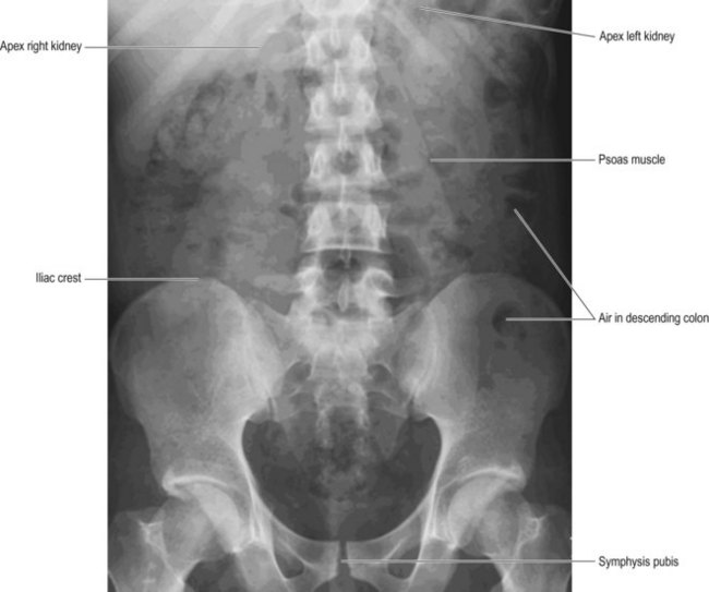
Supine abdomen (Fig. 24.1)
Image receptor (IR) is horizontal, used with antiscatter grid
Positioning
• The patient is supine with the arms slightly abducted from the trunk
• The median sagittal plane (MSP) is coincident with the long axis of the table and the centre of the bucky
• Lead rubber or lead gonad protection is applied, below the symphysis pubis, to male gonads
• Anterior superior iliac spines (ASISs) are equidistant from the table-top
• Because of magnification, owing to the significant distance from the symphysis pubis to the IR, the symphysis pubis should lie well above the lower boundary of the IR
• Central ray and the middle of IR should be accurately aligned
Centring
Positioning for (a) over a point in the midline of the abdomen, level with the iliac crests
Positioning for (b) is to the centre of the IR
Comments on centring, collimation and area of interest
It has been stated that the 11th thoracic vertebra should be included in the collimated field as it lies above the renal outlines and at the tip of the right lower lobe of liver and spleen.1 It is noted that in most adults this would not usually allow for the inclusion of all the upper abdominal contents; however, with the exception of examination of the upper gastrointestinal tract, ultrasound is the most appropriate imaging modality for the upper abdomen. This would negate the necessity for the inclusion of abdominal tissue immediately below the diaphragm. When the supine abdomen position is used to demonstrate the kidneys, ureter and bladder, and additional abdominal information is not required, lateral collimation can be made to the ASIS on each side to more effectively reduce radiation dose.
Traditionally a specific centring point has been given when describing the anteroposterior (AP) supine abdomen. This has usually been stated as level with the iliac crests in the midline, as in centring (a), above.2 Unfortunately, the continuing trend for an increase in average height, noted especially in Europe and the Western world and estimated to be increasing by between 10 and 30 mm per decade,3 affects the amount of body tissue that can now realistically be included on the image. Although the iliac crests do appear midway between the diaphragm and symphysis pubis on the image, centring point (a) will only be useful in smaller patients, i.e. those whose abdominal tissue will actually ‘fit’ within the maximum receptor length. Selection of the centring point/positioning method will therefore depend upon the radiographer’s assessment of the patient’s size.
Stay updated, free articles. Join our Telegram channel

Full access? Get Clinical Tree


