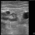
Figure. 10.1
Appendicitis. Acute appendicitis, as identified in this image, can be identified as an aperistaltic, noncompressible, blind-ended tubular structure

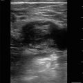
Figure 10.2
Appendicitis with diameter measuring >6 mm. A diameter greater than 6 mm is indicative of acute appendicitis

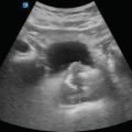
Figure 10.3
Target sign. In short axis, acute appendicitis will often look similar to a target thereby earning the classic finding of “target sign”

Figure 10.4
Appendicolith. Within the inflamed appendix may be a fecalith which is visualized as a hyperechoic rounded structure with posterior acoustic shadowing

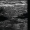
Figure 10.5
Appendicitis with surrounding fluid. Another ultrasonographic finding of acute appendicitis is periappendiceal fluid (arrow)

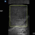
Figure 10.6
Small bowel obstruction. Small bowel obstruction can be identified by dilated, noncompressible small bowel that is greater than 2.5 cm in diameter. There will typically be echogenic material within the lumen of the bowel exhibits and to-and-fro whirling pattern

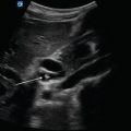
Figure 10.7


Keyboard sign. Keyboard sign can be seen with small bowel obstruction due to prominent plicae circulares, representing piano keys, silhouetted against small bowel contents
Related posts:
Stay updated, free articles. Join our Telegram channel
Full access? Get Clinical Tree
