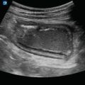
Figure 8.1
Gallbladder in long axis: Gallbladder imaged in long axis

Figure 8.2
Gallbladder in short axis: Gallbladder imaged in short axis

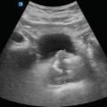
Figure 8.3
Gallbladder wall measurement in short axis: The gallbladder wall should ideally be measured at the thickest location of the anterior wall when in short axis

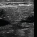
Figure 8.4
Wall measurement in long axis: If unable to obtain an anterior wall measurement, measure the gallbladder wall that abuts against the liver in long axis at the thickest location

Figure 8.5
Portal vein: The portal vein (A) has a thick hyperechoic wall (arrows), which makes it easy to identify within the liver

Figure 8.6
CBD: Common bile duct (arrow) lies anterior to the portal vein

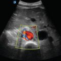
Figure 8.7
Portal triad with color Doppler: Color Doppler can assist in locating the CBD. The portal vein and hepatic artery will both demonstrate color flow, whereas the common bile duct will not

Figure 8.8
Mickey Mouse sign: In cross section, the portal triad will appear as three hypoechoic circles, referred to as mickle mouse sign: common bile duct (A), hepatic artery (B) and the portal vein (C)

Figure 8.9
Normal CBD measurement: Measure the common bile duct should be measured from inner wall to inner wall as pictured here
Gallbladder Pathology
- (a)
Cholelithiasis
Cholelithiasis is the presence of stones within the gallbladder.
Stones appear as hyperechoic, round or oval shaped structures within the gallbladder and exhibit almost complete posterior acoustic shadowing:
Gallstones are usually mobile, such that rolling the patient will cause the stones to move within the gallbladder.
Wall echo shadow (WES) sign is a specific sign that indicates a contracted gallbladder filled with multiple stones [2] or a single large stone [6]:
It is often difficult to appreciate, as the shadowing from multiple stones is often confused with bowel gas shadowing [7, 8]:
Gallstones will produce clean shadowing, whereas bowel gas will produce irregular shadowing [8].
This finding will also preclude visualization of the gallbladder anatomy:
Figure 8.12—Wall echo shadow sign in short axis.
Figure 8.13—Wall echo shadow sign in long axis.
Video 8.7—Wall echo shadow sign.
- (b)
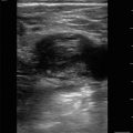
Biliary Sludge
Particulate solids that have precipitated from bile but have not formed into stones.
Sludge will appear as hyperechoic fluid layering within the gallbladder in a gravity dependent fashion.
Sludge will move when the patient is rotated.
Figure 8.14—Sludge.
Video 8.8—Biliary sludge.
- (c)
Get Clinical Tree app for offline access
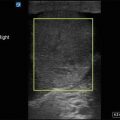
Acute Cholecystitis
Inflammation of the gallbladder.
Findings on ultrasound consistent with acute cholecystitis include thickened gallbladder wall, pericholecystic fluid, and sonographic Murphy’s sign [2]:
Video 8.9—Acute cholecystitis
Thickened gallbladder wall:
Pericholecystic fluid:
Fluid surrounding the gallbladder that develops secondary to inflammationRelated posts:

Stay updated, free articles. Join our Telegram channel

Full access? Get Clinical Tree