27 Abnormal Situs and Cardiac Malposition
Definition and Classification
 Normally, body organs show asymmetric arrangement (Table 27.1). Situs refers to the pattern of arrangement of the body organs relative to the midline.
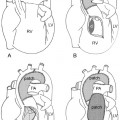
 Situs of the abdominal organs, bronchopulmonary system, and atria should be designated separately. The types of situs at three levels are concordant in the majority of cases (visceroatrial concordance rule), but there are rare exceptions (Fig. 27.1).
 Right isomerism refers to bilateral right-sided anatomy and is associated with asplenia.
 Left isomerism refers to bilateral left-sided anatomy and is almost always associated with polysplenia.
 Cardiac position is independent of body situs. Any cardiac position can be associated with any type of body situs.
 Cardiac malposition is defined as any combination of situs and cardiac position other than a combination of situs solitus and levocardia.
 Incidence of congenital heart diseases according to types of situs and position of the heart:
| Right | Left | |
| Abdominal organs | • Larger lobe of the liver | • Spleen and stomach |
| Lung lobation | • Three lobes | • Two lobes |
| Main bronchus | • Short, eparterial | • Long, hyparterial |
| Pulmonary artery | • Transverse course in front of the right main bronchus | • Oblique course crossing over the left main bronchus (epbronchial course) |
| • Origin of the first branch proximally in the mediastinum | • Origin of the first branch distally at the hilum | |
| Atrium | • Triangular appendage with wide junction demarcated by crista terminalis | • Finger-like appendage with narrow junction not demarcated by crista terminalis |
| • Pectinate muscles extending to the atrioventricular junction | • Pectinate muscles confined to the appendage | |
| • Fossa ovalis with limbus | • No fossa ovalis |
Clinical Manifestations
 Asplenia syndrome with right isomerism:
- • 100% incidence of congenital heart disease, almost always cyanotic heart disease, which presents early in the neonatal period
- • Majority show single-ventricle physiology with pulmonary stenosis or atresia
- • Worst outcome with early death when associated with obstructive type of total anomalous pulmonary venous connection
- Increased risk of serious infections including meningitis and sepsis
- • 100% incidence of congenital heart disease, almost always cyanotic heart disease, which presents early in the neonatal period
 Polysplenia syndromes with left isomerism:
- • Almost always (but not always) associated with congenital heart disease
- • More variable presentation related to the congenital heart disease; most frequently acyanotic heart disease
- • 25% show single-ventricle physiology
- • May present with congestive heart failure due to left-to-right shunt with or without left-sided obstructive lesion, atrioventricular valve regurgitation, or bradycardia
- • Bradycardia with atrioventricular dissociation is common; high mortality in fetal life due to heart block
- • Splenic dysfunction is common despite the presence of multiple spleens.
- • Almost always (but not always) associated with congenital heart disease
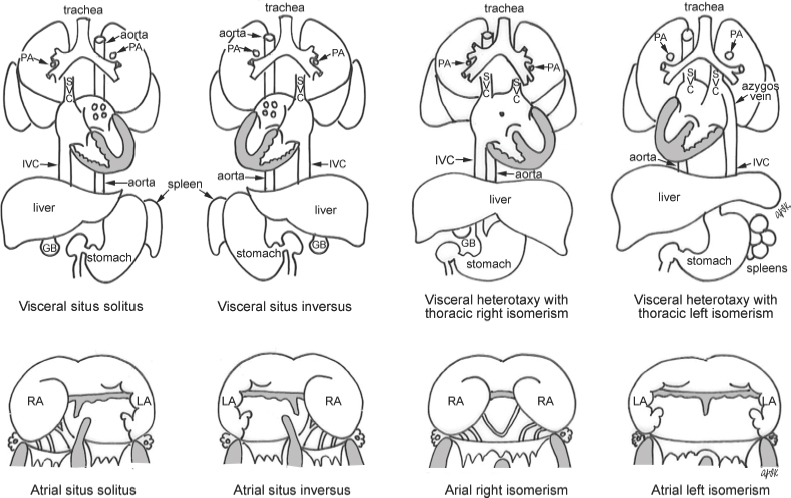
Fig. 27.1 Types of visceral and atrial situs; GB, gallbladder; IVC, inferior vena cava; LA, left atrium; PA, pulmonary artery; RA, right atrium; SVC, superior vena cava.
| Right Isomerism | Left Isomerism | |
| Bilateral superior venae cavae | 45% | 45% |
| Bilateral systemic venous drainage | 70% | 60% |
| Absence of coronary sinus | ~100% | ~60% |
| Interruption of the inferior vena cava | <2.5% | 80% |
| Juxtaposition of the aorta and inferior vena cava | ~90% | Uncommon |
| Extracardiac type of total anomalous pulmonary venous connection with/without obstruction | 50%, with obstruction in 50% | Rare |
| Pulmonary venous connection to ipsilateral atriums | 4% | 45% |
| Atrioventricular septal defect | 90% | 50% |
| Atrial septum Functionally common atrium in 50% |