Acute Arterial Occlusive Disease of the Upper Extremity


Acute Arterial Occlusive Disease of the Upper Extremity


Jeff Dai-Chee Tam, Mark F. Given, Stuart M. Lyon and Kenneth R. Thomson


In the absence of trauma, acute upper limb arterial disease is relatively uncommon, particularly in comparison with lower limb disease. Some authors suggest that it is one sixth as common as that in the leg.1 The majority of patients will have small vessel disease.2 Blunt trauma and repetitive use of the arm—for example, in athletes or associated with certain occupations—are the usual causes in the proximal upper extremity.3,4 More distal upper limb ischemia is usually the result of emboli from a proximal stenosis or a cardiac origin. Occlusive disease of the upper extremity is as important as lower limb occlusive disease, not least because it may reflect an underlying systemic illness.5,6


Atherosclerotic disease is the most common cause of large vessel stenosis or occlusion, but it may also present as small vessel occlusion via thromboembolism. Aneurysmal disease, including cystic adventitial disease, may also be a source of emboli.7


Other primary causes include thoracic outlet compression syndrome, drug-induced occlusion, post-radiotherapy stenoses, and iatrogenic catheter injury.5,8,9


Technique


Anatomy and Approaches


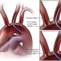
The arterial supply to the upper limb originates from the subclavian arteries. The left subclavian artery arises directly from the aortic arch in the majority of cases, whereas the right subclavian originates from the brachiocephalic artery. The subclavian vessels first pass between the scalene muscles and then between the clavicle and first rib. At the lateral border of the clavicle they become the axillary arteries.


The branches of the subclavian artery include the vertebral, internal mammary, thyrocervical trunk, costocervical trunk, and supreme intercostal arteries.


The axillary artery extends to the lateral margin of teres minor, where it becomes the brachial artery. Its branches include the superior and lateral thoracic arteries and the thoracoacromial, subscapular, scapular circumflex, and circumflex humeral arteries. The radial, ulnar, and median nerves are contained in a connective tissue sheath along with this vessel and are at risk of damage if one is considering puncture of an axillary artery. Also of importance is that there are multiple potential collateral pathways involving the branches of both the subclavian and axillary arteries in the presence of stenosis or occlusion.


Lateral to the teres minor, the axillary artery becomes the brachial artery, which has the profunda brachialis as its first branch in the upper arm. This branch travels posterolaterally around the humerus, giving off multiple muscular branches and providing collateral branches around the elbow.


At the elbow joint, the brachial artery then terminates into the radial, ulnar, and interosseus arteries. The interosseus artery then divides into an anterior and posterior branch. The radial and ulnar arteries continue down the forearm to form the superficial and deep palmar arches and supply the hand. The deep palmar arch is more proximal and is mainly supplied by the radial artery, whereas the superficial arch is more distal and has the ulnar artery as its main supply. These classic palmar arcades occur in fewer than 50% of patients, but there are multiple variations. The classic anatomic description of the more proximal upper limb arteries noted earlier is also subject to variation, particularly in the origin of their branches, and this can be a potential source of confusion on angiography.

Stay updated, free articles. Join our Telegram channel

Dec 23, 2015 | Posted by in INTERVENTIONAL RADIOLOGY | Comments Off on Acute Arterial Occlusive Disease of the Upper Extremity

Full access? Get Clinical Tree

Get Clinical Tree app for offline access