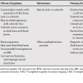
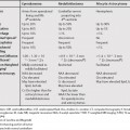
91 Adrenal adenomas are the most common adrenal lesions. As an incidental finding, a small homogeneous adrenal lesion is highly likely to be an adenoma. In patients with cancer, it is often important to determine whether an adrenal lesion is an adenoma or metastasis. The methods below should be only applied to relatively homogeneous adrenal lesions. Adrenal adenomas generally cannot be diagnosed by imaging in masses with substantial areas of necrosis or hemorrhage.1 Adrenal adenomas have intracytoplasmic lipid that is not present in metastases. Detection of lipid content differentiates adenomas from metastases. This can be evaluated by noncontrast computed tomography (CT) or chemical shift magnetic resonance imaging (MRI). Adenomas have lower attenuation values than metastases. Using a threshold attenuation value of 10 Hounsfield units (HU), the sensitivity is 71% and the specificity 98%.2 Adenomas demonstrate signal loss on opposed phased chemical shift imaging. Both visual and quantitative analysis can be used to evaluate whether there is signal loss. Two quantitative parameters that have been used to diagnose lipid content are Ten to 40% of adrenal adenomas will be lipid poor4 and cannot be diagnosed as adenomas on noncontrast CT. Although adenomas can have noncontrast CT attenuation values >10, a non-calcified nonhemorrhagic adrenal lesion measuring >43 HU is suspicious for malignancy.5 In lesions with an attenuation value >10 but <43 on noncontrast CT, chemical shift MRI and contrast-enhanced CT may be helpful for diagnosis.
Adrenal Adenoma versus Metastasis
Diagnosis of Lipid-Rich Adrenal Adenomas
Noncontrast Computed Tomography
Chemical Shift Magnetic Resonance Imaging
Diagnosis of Lipid-Poor Adrenal Adenomas
Chemical Shift Magnetic Resonance Imaging
Stay updated, free articles. Join our Telegram channel

Full access? Get Clinical Tree


