Adrenal Venous Sampling


Adrenal Venous Sampling


Kenneth R. Thomson, Jim Koukounaras and Mark F. Given


In the investigation of adrenocortical function, urine and blood cortisol measurements as well as adrenocorticotropic hormone (ACTH, corticotropin) levels are used. Adrenal venous sampling is almost never required in the investigation of underproduction (Addison disease) or overproduction (Cushing disease) of cortisol. These can be diagnosed by peripheral samples during an ACTH stimulation test. Levels less than 2 µg/dL indicate Addison disease and greater than 22 µg/dL indicate Cushing disease.


Excessive excretion of aldosterone may be primary due to a tumor (Conn syndrome) or hyperactivity of the outer area of the adrenal cortex, or secondary due to nonadrenal conditions that cause a severe imbalance in sodium and potassium. Examples of secondary causes of hyperaldosteronism are congestive heart failure, cirrhosis with ascites, depletion of sodium from diuretics, or toxemia of pregnancy. Plasma renin activity is typically elevated in secondary hyperaldosteronism and reduced in primary aldosteronism. As a result, aldosterone and renin are usually measured together.


Primary aldosteronism is increasingly considered to be a remedial cause of hypertension. Patients with an increased peripheral aldosterone/renin ratio after correction for hypokalemia and removal of interfering medication who also show a positive response to corticosteroid suppression should be considered for adrenal vein sampling to distinguish unilateral from bilateral primary aldosteronism. Because corticosteroid suppression requires sodium loading, it is generally not performed in the elderly or those with severe hypertension. It has been suggested that a saline infusion test is equally reliable, but it is limited in persons with heart failure. In most cases, a prior computed tomography (CT) scan of the adrenal glands will have been performed. If there is evidence of a unilateral enlargement of a portion of the adrenal gland, there is slightly more likelihood of unilateral aldosteronism excess. In about 40% of cases of aldosteronism, however, the CT is unhelpful or even misleading.


Technique


Anatomy and Approach


The adrenal gland is a composite retroperitoneal organ with a medulla of ectodermal origin and a cortex of mesodermal origin. The gland shrinks significantly in the first 2 weeks after birth as the fetal cortex degenerates. The adult cortex is fully differentiated by puberty. The cortex forms the main mass (90%) of the gland and is richly supplied by arteries. A single vein emerges from each adrenal gland. The arterial supply is multiple and comes from the aorta directly, the renal artery, and the inferior phrenic artery.


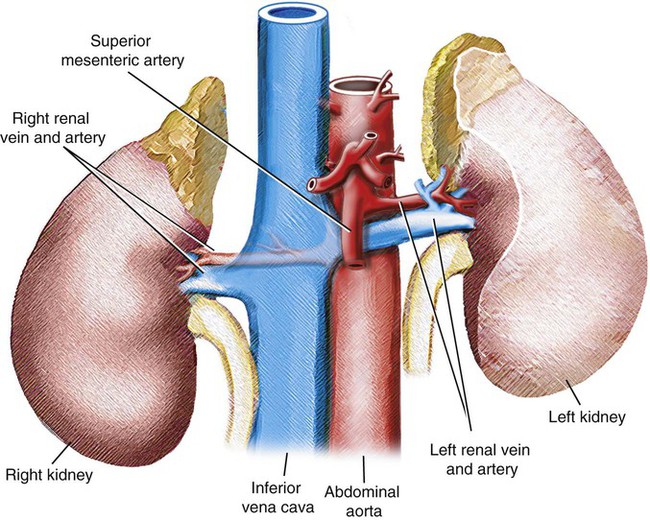
The adrenal glands lie at the anterosuperior aspect of the upper pole of each kidney and are enclosed within the renal fascia but separated from the kidneys by loose fibroareolar tissue (Fig. 109-1). There maybe several small masses of tissue identical with adrenal cortex “cortical bodies” in the neighborhood of the gland.


The right adrenal gland is roughly triangular, with its apex and medial portion of the anterior surface in contact with the posterior aspect of the inferior vena cava (IVC) (Fig. 109-2

Only gold members can continue reading. Log In or Register to continue

Stay updated, free articles. Join our Telegram channel

Dec 23, 2015 | Posted by in INTERVENTIONAL RADIOLOGY | Comments Off on Adrenal Venous Sampling

Full access? Get Clinical Tree

Get Clinical Tree app for offline access