Fig. 35.1
Simple radiographic features of multiple myeloma. Lateral skull (a) and pelvis anteroposterior (b) radiographs show multiple variable punched-out osteolytic lesions without definite marginal sclerosis and periosteal reaction
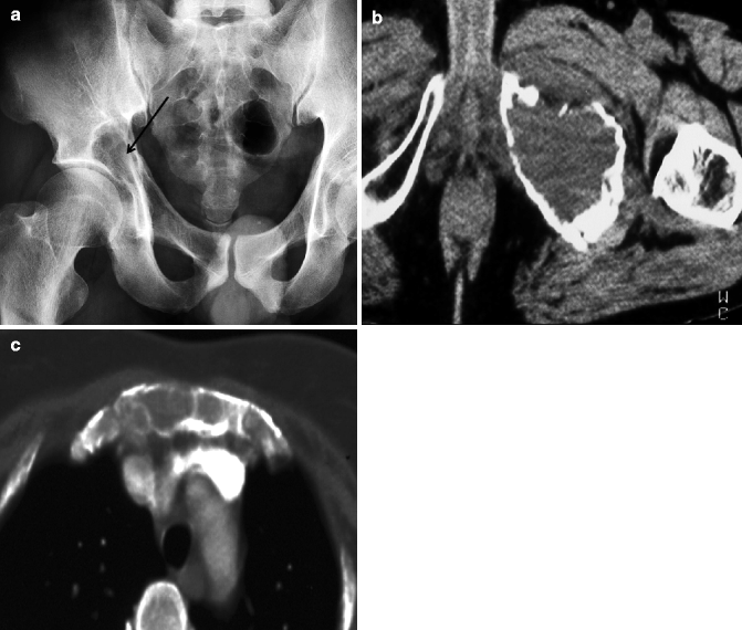
Fig. 35.2
Radiographic features of plasmacytoma. Pelvis anteroposterior view (a) shows an expansile bony destructive lesion in the right acetabulum (arrow). Axial CT of the pelvis and sternum in another patient (b, c) shows bony destruction and endosteal scalloping and fractures in the left pubis and sternum
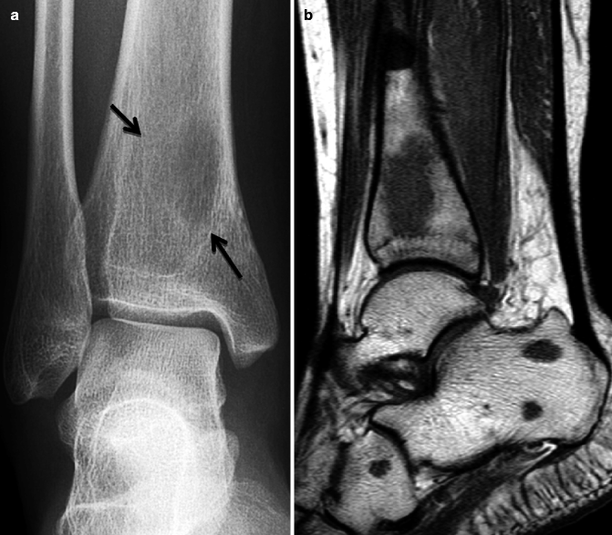
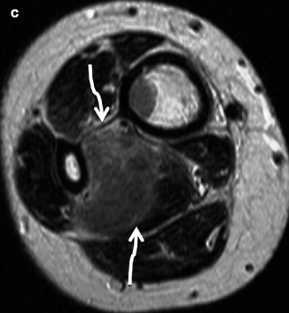
Fig. 35.3
Multiple myeloma involving the distal tibia and surrounding muscle. (a) Simple radiography of the right ankle shows an osteolytic lesion with ill-defined margins (arrows) in the distal tibia. T1-weighted sagittal MRI (b) shows multiple bony destructive lesions with low signal intensity in the tibia, calcaneus, and cuboid. T2-weighted axial image (c) shows mass lesion with high signal intensity in the right flexor hallucis longus muscle (curved arrows)
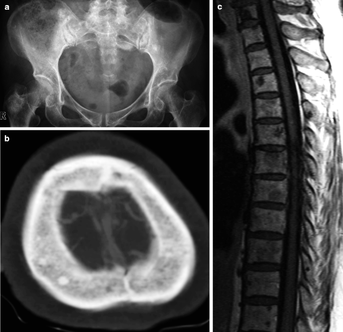
Fig. 35.4
POEMS syndrome with sclerotic lesions. Multiple sclerotic lesions are noted in the right greater trochanter, left iliac bone (a), and skull (b). T1-weighted image (c) shows multifocal lesions with variable-sized low signal intensities
Radiography is known to be more sensitive than bone scans in myeloma because osteoblastic activity is very low relative to the degree of bone destruction. Only about 10–25 % cases are positive on bone scan. The most sensitive imaging modality is MRI which can show direct bone marrow invasion. On T1-weighted imaging, focal or diffuse lesions clearly contrast with the homogeneous high signal intensity of normal fat marrow in adults. MRI findings of multiple myeloma occur in three broad patterns: focal, variegated, and diffuse (Moulopoulos et al. 1992). The focal pattern lesion is most common and it is often equivalent to bone destruction on radiography (Fig. 35.5). This lesion is hypointense on T1-weighted imaging and hyperintense on T2-weighted imaging against the background of normal bone marrow and generally strongly enhances following contrast. However, the lesion can be hypointense on both T1-weighted imaging and T2-weighted imaging and occasionally shows poor contrast enhancement. Approximately 25 % of multiple myeloma presents as a diffuse homogenous lesion which is difficult to detect on radiography and has a poor prognosis. In addition, MRI findings can be used to predict the progress of the disease. Disease progress is more rapid in patients with the diffuse pattern than in subjects with the variegated or focal pattern. Response to treatment is also apparent on MR investigation (Moulopoulos et al. 1994). Complete remission demonstrates no contrast enhancement or peripheral enhancement within the bone marrow abnormality. On the other hand, partial remission shows residual contrast enhancement while in some cases a diffuse pattern may change into focal or variegated patterns. One fundamental role of MRI in multiple myeloma is to evaluate patients with neurological symptoms, because immediate treatment is necessary when a focal vertebral lesion compresses the spinal cord.
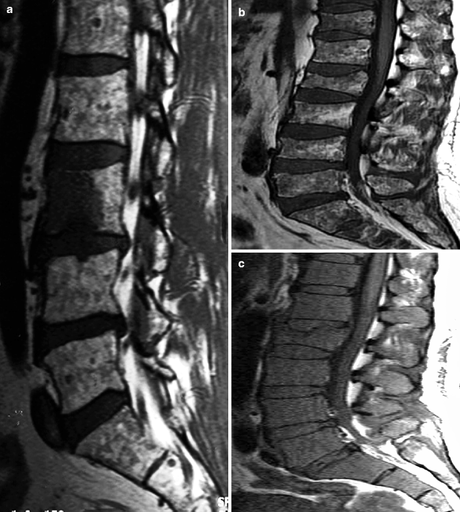

Fig. 35.5
Various patterns of the bone marrow invasion in multiple myeloma on T1-weighted images. (a) Focal mass formation in the spine (focal form), (b) small speckled low-density lesions of the bone marrow (variegated form), and (c) homogenous low signal intensity of the bone marrow (diffuse form)
Radiography cannot detect bone density loss until the loss reaches 30–50 %; and thus, radiography is impractical for detecting this disease. Technical developments in MRI have made the staging process of multiple myeloma faster and more accurate than radiography (Ghanem et al. 2006). The detecting rate for FDG PET-CT is similar to MRI in multiple myeloma of the vertebrae and pelvic bone. FDG PET has the advantage of enabling assessment of the whole body for detection and staging of multiple myeloma (Fig. 35.6). PET-CT is known to be more effective than whole-body MRI in the evaluation of treatment response even though its high sensitivity can overstage the disease (Lutje et al. 2009).
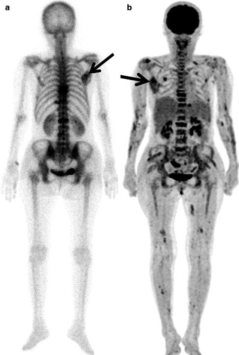
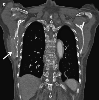


Fig. 35.6
Multiple myeloma in a 65-year-old woman. Bone scan (a), MIP (maximum intensity projection) image of PET-CT (b), and coronal CT (c) taken within 1 week show multiple lesions in the spine and the ribs as well as in the right scapula (arrow). PET-CT depicts more bone lesions than a bone scan with high FDG uptake (mSUV: 8.6)
35.4 Amyloidosis
Amyloidosis is characterized by deposition of amyloid protein, which is a glycoprotein not normally present in that tissue or organ. Systemic amyloidosis may occur on its own as a primary event or secondary to chronic inflammation, tuberculosis, multiple myeloma, rheumatoid arthritis, or connective tissue disease. Focal involvement or localized amyloidosis includes gingival and tongue deposition leading to functional disability. In addition, beta-2-microglobulin amyloidosis can occur in patients undergoing long-term hemodialysis. The diagnostic histological test shows an amorphous eosinophilic appearance on hematoxylin and eosin stain, while Congo red and polarized light (green) can be used for confirmation. Amyloidosis can involve various regions such as the shoulder, hip, and knee joints and may cause back pain with limitation of spinal motion. Deposition of amyloid in the soft tissue can cause compression symptoms such as carpal tunnel syndrome.
Imaging findings of amyloidosis include soft tissue mass from deposition in the bone with well-marginated bony erosions, subchondral bone cysts, or decreased bone density and deposition in the synovial membrane leading to widening of the joint space, joint contracture, and atrophy of muscle.
Joint changes may be rather similar to the findings of rheumatoid arthritis (Katoh et al. 2008), but amyloid arthropathy can be distinguished by the presence of a large soft tissue mass, the lack of joint space narrowing, and the presence of well-marginated subchondral bone cysts and erosions. A differential diagnosis includes gouty arthritis, xanthomatosis, and pigmented villonodular synovitis. Deposition of amyloid around the blood vessels can cause osteonecrosis of the epiphyseal regions. Amyloid deposition on MRI is seen as soft tissue with signal intensity between that of muscle and fibrocartilage (Fukusa and Yamamoto 2001) with additional features such as thickening of the synovium, paratendinous deposition, joint effusion, and bone erosion (Fig. 35.7).
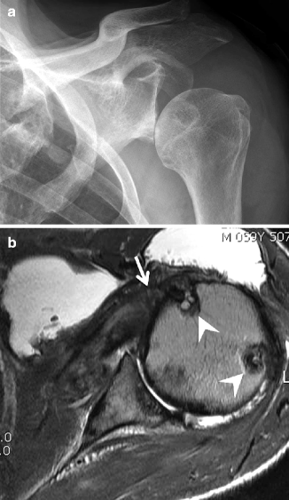

Fig. 35.7
Amyloidosis of the left shoulder in a 59-year-old man. Oblique simple radiography (a) shows increased density and downward displacement of the humeral head by the soft tissue mass density. (b) Fat suppressed axial T1-weighted MR arthrography shows thickening of the rotator cuff (arrow) and multiple bony erosions (arrowheads)
35.5 Leukemia
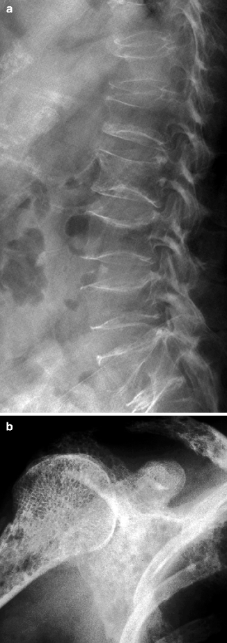
Leukemia is a malignancy of myelogenous or lymphocytic white cells which can occur in both acute and chronic forms. Leukemia is one of the most common cancers of children; 75 % of leukemias are acute lymphoblastic leukemia (ALL), 15–20 % are acute myeloid leukemia (AML), and 5 % are chronic myeloid leukemia (CML). Clinical manifestations include focal or diffuse bone pain or joint pain, joint effusion, fever, anemia, and recurrent infection. Radiographic features are normal or decreased bone density with a permeative pattern or vertebral compression fracture secondary to leukemic infiltration (Fig. 35.8). A transverse radiolucent line through the metaphyses termed a leukemic line can be found around the knee joint, the ends of the humerus, or even in the vertebral body. Leukemic line is due to impaired bone formation rather than direct infiltration of leukemia cells and can be also be found in children under 2 years of age with severe malnutrition or systemic disease. These transverse lines transform into sclerotic lines after treatment. In some cases, there are multiple well-marginated lytic lesions and punched-out lesions, but osteoblastic lesions are extremely rare. Occasionally a laminar or sunburst type of periosteal reaction can be present. MRI (Olson et al. 1986) is more sensitive at detecting bone marrow lesions of leukemia patients. Most patients have a diffuse bone marrow infiltration pattern on MRI (Fig. 35.9), although various heterogeneous patterns may be seen depending on the degree of marrow cellularity. Histological examination is necessary to confirm the diagnosis and to subtype the leukemic process, while repeated bone marrow biopsy is needed to evaluate treatment response. The role of MRI of the bone marrow has been studied as an alternative to invasive bone biopsy. An increased fat ratio is indicative of a good treatment response (Vande Berg et al. 1996). However, elimination of leukemic cells and regeneration of bone marrow occur independently, and changes in T1 relaxation time are not a sensitive indicator of treatment response. Dynamic contrast-enhanced MRI is expected to provide practical information on treatment response since the time and degree of contrast enhancement vary with the cellularity and perfusion of the bone marrow.
Get Clinical Tree app for offline access

Fig. 35.8
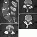
Acute lymphoblastic leukemia in an elderly patient. (a) Lateral radiography shows osteopenia and multiple insufficiency fractures with flat and biconcave patterns. (b


Stay updated, free articles. Join our Telegram channel

Full access? Get Clinical Tree


