Fig. 41.1
Diagram illustrates the relationship between the vertebral bodies and the intervertebral disc, as well as the different components and internal structure of the disc. The intervertebral disc consists of a central nucleus pulposus surrounded by the annulus fibrosis and cartilaginous vertebral end plates (Reproduced with permission from Peh (2009))
The innervation of the intervertebral disc is via two interconnected nerve plexuses supplying the anterior and posterior parts of the disc. These receive components from branches of two sympathetic trunks, the proximal ends of the gray rami and the perivascular nerve plexuses of the segmental arteries, as well as the sinuvertebral nerve, which is the primary contributor to the posterior plexus. Nerve endings are found in the periphery of the outer annulus at a depth of a few millimeters in a normal disc, with the highest concentration in the posterolateral aspects of the annulus fibrosis. Deeper penetration of nerve endings has been shown in discs that are degenerate (Walker et al. 2008).
41.3 Pathophysiology
The pathophysiology of discogenic pain is incompletely understood. Internal disc disruption has been postulated as the root cause. The characteristic feature of internal disc disruption is a radial fissure extending to the innervated outer third of the annulus fibrosis, causing irritation of the nerve endings and provoking pain. This may occur in the absence of disc protrusion. A torn portion of the annulus fibrosis may result in abnormal stress distribution to the rest of the fibers, causing increased pressure and lowering the threshold for pain stimulation. Chemical factors have also been implicated. Structural damage in the disc and disruption of the annulus fibrosis release chemical substances such as phospholipase A2 and other cytokines, including tumor necrosis factor into the spinal canal, which provoke spinal pain (Saal et al. 1990; Spiliopoulou et al. 1994; Roberts et al. 1995; Coppes et al. 1997; Walker et al. 2008). As the annulus fibrosis weakens and tears, part of the nucleus pulposus herniates through the defect into the spinal canal and neural foramen, impinging on an adjacent nerve root and causing typical radicular pain. However, nerve root compression is the cause of pain in only 5 % or fewer of the patients with degenerative disc disease (Tehranzadeh 1998).
41.4 Indications and Contraindications
Discography, being an invasive procedure, involves small but definite risks and should not be used as a screening study for back pain. Careful selection of patients with appropriate indications is paramount to avoid unnecessary procedures and to maximize patient benefit. In general, the patient should have persistent back pain for duration of at least 4 months, which is not responding to conservative treatment and for which surgery is considered. Discography should be considered only if MRI has failed to reveal the etiology of back pain. Noninvasive tests such as radiographs, CT, and MRI should be employed to exclude other non-discogenic causes of back pain such as facetogenic, neoplastic, inflammatory, and traumatic causes (Peh 2005, 2009).
41.4.1 Indications
Specific indications for provocative discography include the following (Guyer and Ohnmeiss 1995; Bini et al. 2002; Fenton and Czervionke 2003; Guyer et al. 2003; Anderson 2004; Peh 2005):
Evaluation of an abnormal disc detected on other imaging modalities to assess its full extent or for correlation with clinical symptoms
Investigation of persistent, severe symptoms that do not correlate with MRI or CT findings
Identification of specific disc levels which are symptomatic in cases where MRI or CT shows multilevel disease, to facilitate surgical planning
Assessment prior to vertebral fusion to determine if the intervening disc is symptomatic and to assess the adjacent discs
Assessment of the disc prior to percutaneously directed therapies such as intradiscal electrothermal therapy (Saal and Saal 2000; Wetzel et al. 2002; Davis et al. 2004; Pauza et al. 2004)
Assessment of patients prior to minimally invasive surgery to confirm that disc herniation is contained, or to investigate contrast distribution before chemonucleolysis
Detection of recurrent disc herniation in a postsurgical patient with failed back syndrome in whom MRI is nondiagnostic
41.4.2 Contraindications
Contraindications include (Tehranzadeh 1998; Fenton and Czervionke 2003; Anderson 2004; Peh 2005):
Pregnancy
Systemic infection or skin infection over the puncture site
Severe allergy to injectate, especially the contrast agent
Previously operated disc
Solid bone fusion that does not allow access to the disc
Severe spinal cord compromise at the disc level to be investigated
41.5 Technique
Since its initial description more than six decades ago, discography has evolved with many refinements to the technique, resulting in improved sensitivity and specificity. The detection of morphologic abnormalities on discography is becoming less relevant in the current age of high-resolution and readily available MRI. This puts the provocative aspect of discography in the spotlight as a diagnostic aid in clinical decision-making. When performed appropriately, discography can enhance sensitivity and specificity compared to non-provocative imaging (Manchikanti et al. 2009b).
41.5.1 Pre-procedure Assessment
The patient should be interviewed about the type, location, and nature of the pain and any history of prior spinal surgery. Informed consent obtained prior to the procedure would include discussion regarding the nature and purpose of the pain provocation test as well as the possible risks and complications. Drug and contrast allergies should be elicited and addressed accordingly. MR discography with gadolinium contrast is an option for patients with severe iodine contrast allergy and has been shown to be accurate and safe (Huang et al. 2002; Slipman et al. 2002; Falco and Moran 2003; Streitparth et al. 2011). The patient’s medical records and prior imaging studies should be reviewed. Previous MRI studies are valuable for assessing overall disc morphology and for identification of a normal disc that can be used as a control. Careful pre-procedure evaluation with radiographs as well as MRI may be helpful to avoid ambiguity regarding transitional lumbar anatomy. Fasting for 6 h prior to the procedure is recommended (Peh 2005). Anticoagulant and antiplatelet medications such as warfarin and aspirin are withheld 5 days prior to the procedure to avoid possible bleeding. Pain medications, anti-inflammatory drugs, sedatives, and any medications that may alter the patient’s perception to pain should not be used on the day of the procedure to ensure integrity of test results. Some authors prescribe prophylactic intravenous antibiotics (cefazolin 1 g bolus or clindamycin 600 mg if allergic to cephalosporin or penicillin) to be administered within 1 h before commencement of the procedure. Blood pressure and pulse oximetry should be monitored throughout the procedure. A strictly aseptic technique is employed (Walker et al. 2008).
41.5.2 Equipment
Provocative discography is best performed in an interventional suite within the diagnostic radiology department using a biplane fluoroscope or high-quality C-arm fluoroscope as an acceptable alternative if the prior is unavailable. For patients who are allergic to iodinated contrast agents, MR discography using intradiscal gadolinium-chelate is a viable alternative (Huang et al. 2002; Slipman et al. 2002; Falco and Moran 2003; Streitparth et al. 2011). There are variations in the size and type of needles used by different centers and practitioners. Some practitioners advocate the single-needle approach using a styleted needle that ranges in size from 18 to 22 gauge (Fenton and Czervionke 2003). Many practitioners adopt the double-needle approach which has been shown to result in a lower rate of discitis as the inner needle which enters the nucleus pulposus does not come in contact with the skin (Fraser et al. 1987; Peh 2005). The use of a thinner 26-gauge inner needle also decreases the size of the puncture hole in the annulus fibrosis, and having a pre-shaped curve at the distal end of the inner needle facilitates entry into center of the L5–S1 nucleus.
An example of a discography set consists of the following (Peh 2005):
21-gauge 12.5 cm long stainless steel spinal needle with stylet and a 26-gauge 16.0 cm long stainless steel spinal needle with stylet for thoracic and lumbar discography.
A 20-gauge 6.35 cm long stainless steel spinal needle with stylet and a 26-gauge 8.9 cm long stainless steel spinal needle with stylet for cervical and thoracic discography.
A curved needle set consisting of a 21-gauge 10.0 cm long stainless steel straight needle with stylet and a 26-gauge 15.0 cm long nitinol curved needle is preferred for the L5/S1 disc.
25-gauge, 3.8 cm needle for skin and subcutaneous local anesthesia.
1 ml tuberculin syringe and nonionic contrast agent 350 mg I/ml (Omnipaque 350) for intradiscal injection.
5 ml syringe and lidocaine 1 % for local anesthesia.
Alcohol and povidone-iodine scrubs.
Sterile gauze and drapes.
41.5.3 Cervical Discography
Cervical discography is controversial, with some investigators recommending against the procedure as the information obtained from cervical discography does not outweigh the increased risks of complications that are reported to occur in up to 13 % of cases (Connor and Darden 1993). These complications include discitis, epidural abscess, hematoma, myelopathy, and quadriplegia (Zeidman et al. 1995). Other practitioners have found cervical discography to be a safe and useful procedure in selected patients with chronic intractable neck pain with negative or indeterminate imaging findings and who are being considered for surgery (Connor and Darden 1993; Zeidman et al. 1995; Grubb and Kelly 2000). A recent systematic review of cervical discography concluded that cervical discography performed according to the International Association for the Study of Pain (IASP) criteria may be a useful tool for evaluating chronic cervical pain, although not disc herniation or radiculitis. However, the level of evidence is limited by lack of large studies and consistent use of IASP standards (Manchikanti et al. 2009a).
The cervical disc cannot be approached posteriorly because of the spinal cord, anteriorly because of the airway, or posterolaterally because of the vertebral artery. As such, cervical discography is performed with an anterolateral approach with the patient supine and neck slightly hyperextended. A right-sided approach is preferred as most operators are right-handed, and it also decreases the likelihood of inadvertent puncture of the esophagus which lies slightly to the left. Careful technique and accurate surface localization are important to avoid injury to vital structures in the neck. The carotid sheath contents are protected by palpating the carotid pulse and displacing the carotid sheath laterally with the middle and index fingers. A 20-gauge 6.35 cm outer needle is introduced just medial to the fingertips following local anesthetic administration, at an angle of 40–45°, taking care to avoid the esophagus and trachea (Fig. 41.2). Under intermittent fluoroscopy, the needle tip is then slowly directed to the outer annulus fibrosis of the disc where it should be positioned in line with the anterior cortex of adjacent vertebral bodies on lateral projection and the ipsilateral pedicles on anteroposterior projection. The stylet is then removed and replaced by 26-gauge, 8.9 cm inner needle which is directed to the center of the disc space under fluoroscopy (Fig. 41.3) (Peh 2005).
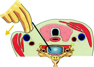
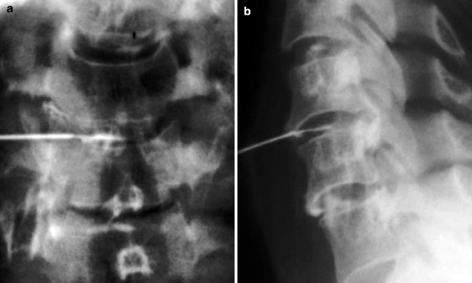
Fig. 41.2
Needle insertion for cervical discography. The carotid sheath contents are protected by lateral displacement using the fingers. Care is taken to avoid other structures such as the trachea and esophagus by angling the needle at 40 to 45° (Reproduced with permission from Peh (2009))
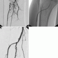
Fig. 41.3
Needle positions for cervical discography. (a) Anteroposterior and (b) lateral radiographs show positions of the inner and outer needles at the C4/5 disc (Reproduced with permission from Peh (2009))
41.5.4 Thoracic Discography
There are very few indications for thoracic discography which is rarely performed or reported in the literature. Severe and disabling thoracic pain secondary to disc degeneration that requires discography has not been well studied (Tehranzadeh 1998; Fenton and Czervionke 2003). This procedure has been used to evaluate symptomatic Scheuermann disease (Winter and Schellhas 1996). Thoracic discs with prominent Schmorl nodes may be intensely painful on provocation, even in asymptomatic subjects, and thoracic discography may demonstrate disc pathology that is not seen on MRI (Wood et al. 1999). The technique is similar to lumbar discography described below, with selection of needle length based on the depth of the disc to be examined. The needle tip should be kept along the lateral aspect of the superior articular process on posteroanterior fluoroscopy to avoid entering the spinal canal. A pleural puncture is likewise avoided by keeping the needle tip medial to the costotransverse junction (Fenton and Czervionke 2003).
41.5.5 Lumbar Discography
Provocative discography is most commonly performed for evaluation of the lower three lumbar discs. For lumbar discography, the patient may be placed in a prone or left lateral decubitus position, depending on operator preference. Some advocate the prone position in which the patient is more stable and immobile (Tehranzadeh 1998; Fenton and Czervionke 2003). The principal author prefers the left lateral decubitus position with the patient flexing his or her knees and a pillow placed beneath the waist to keep the spine straight. The skin puncture point is approximately 8–10 cm from the midline. After the patient is cleaned and draped, and local anesthesia administered, the outer discography needle is then inserted. The posterolateral extradural approach is preferred as it avoids puncturing the thecal sac (Tehranzadeh 1998; Fenton and Czervionke 2003; Anderson 2004). The outer needle is inserted with an obliquity of about 45–60º to the sagittal plane (Fig. 41.4).
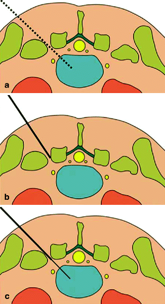
Fig. 41.4
Needle positions during lumbar discography. (a) The dotted line represents the planned needle path. (b) The position of the outer needle is directed towards the posterolateral corner of the disc. (c) The inner needle is inserted within the outer needle and directed to the center of the nucleus pulposus (Reproduced with permission from Peh (2009))
Under intermittent fluoroscopic imaging in the AP and lateral directions, the outer needle is positioned so that its tip is placed at the right posterolateral corner of the annulus fibrosis of the target disc. The needle tip should lie in line with the posterior cortex of the adjacent vertebral bodies on the lateral projection and with the ipsilateral pedicles of the adjacent vertebral bodies on the anteroposterior projection (Fig. 41.5). For the L5–S1 disc, an additional caudal angulation of up to 40º is usually necessary, due to the overlying iliac crest. Pre-bending of the inner and outer needle or the use of a curved needle set may be necessary to facilitate entry into the disc in difficult cases at this level. Mild rubbery but firm resistance is felt when the needle tip comes into contact with the annulus fibrosis. The stylet of the outer needle is then removed and replaced by the longer inner needle. Under fluoroscopic guidance in two orthogonal directions, the tip of the inner needle is directed to the center of the nucleus pulposus. When the position of the inner needle is satisfactory, a test injection of 0.1 ml of nonionic contrast agent is made to confirm the needle position using a 1 ml tuberculin syringe with 0.1 ml markings (Peh 2009). If the position of the needle tip is suboptimal, adjustment of needle position and repeat fluoroscopic screening are required.
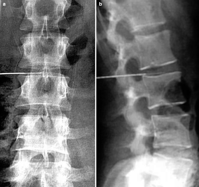
Fig. 41.5
Needle positions for lumbar discography. (a) Anteroposterior and (b) lateral radiographs show needle positions of the outer and inner needles in the L2/3 disc. The tip of the outer needle is in line with the ipsilateral pedicle on the frontal view and the posterior vertebral body cortices on lateral view. The tip of the smaller inner needle is seen near the center of the L2/3 disc
The injected contrast agent should form a rounded or curvilinear blob near the center of the disc space. Care should be taken to avoid injection into the annulus fibrosis which may lead to a false positive response. This is manifested by marked resistance to contrast instillation at the start of the injection and visualization of the contrast agent persisting between the needle tip and the edge of the disc (Peh 2009). In a normal disc, there is moderate resistance during contrast injection up to a volume of 1.5 ml which is the usual capacity of a nondegenerate disc. In a degenerate disc, there may be mild or no resistance to contrast injection with a typical volume of more than 2 ml. The volume of contrast agent injected should not exceed 3 ml for lumbar discography, 0.5 ml for cervical, and 1.0 ml for thoracic discography (Tehranzadeh 1998). The injection is usually terminated when very firm resistance is felt or if severe pain is produced (Fenton and Czervionke 2003). The volume of contrast agent injected at each level as well as the amount of resistance encountered should be documented.
41.5.6 Recovery
Following removal of the needles and ensuring that there is no excessive bleeding, small adhesive bandages applied over the puncture sites are all that is required. After completing post-discography imaging, the patient should be observed for up to 2 h in either a reclining or recumbent position with regular monitoring of vital signs. Upon discharge, most practitioners will give their patients a prescription of a non-narcotic painkiller, with an option of prescribing a short prophylactic course of oral broad-spectrum antibiotics (Fenton and Czervionke 2003; Peh 2005).
41.6 Interpretation
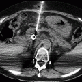
The two major aspects to consider in the interpretation of discography are disc morphology and pain provocation (Peh 2005). Discography interpretation may be supplemented by performing post-procedure imaging using CT. CT discography provides excellent anatomical details in the axial plane and allows for good quality reconstructions in the sagittal or coronal planes when using a multislice scanner, which may provide useful additional information (Vivitmongkonchai and Peh 2001).
41.6.1 Disc Morphology
Disc morphology can be assessed with either radiographs or CT after intradiscal contrast injection. A normal disc maintains a normal height on both anteroposterior and lateral radiographs. The injected contrast agent remains in a central position within the nucleus pulposus, taking on a unilocular (“cotton ball” or rectangular) or bilocular (“hamburger bun”) configuration (Fig. 41.6). A Schmorl node is sometimes seen as focal protrusion of injected contrast agent into the adjacent vertebral end plate (Brightbill et al. 1994). A degenerated disc will show a reduced disc height with irregular fissures in the annulus fibrosis which may be complex or multiple. The peripheral annulus may remain intact or be torn. Contrast extravasation beyond the margins of the disc is indicative of an annular tear. A bulging disc may be seen with an intact peripheral annulus and is characterized by circumferential, diffuse, and symmetrical annular bulging. A disc protrusion refers to focal, often asymmetrical, posterior or posterolateral protrusion of disc material within an intact posterior longitudinal ligament. Superior or inferior migration of the nuclear material may give a “candle drip” appearance. A disc extrusion is a large disc protrusion that involves the posterior longitudinal ligament with contrast agent seen within the epidural space on discography. Extruded disc material in the extradural space which has separated from the parent disc is called a sequestrated disc (Figs. 41.7 and 41.8) (Peh 2005).
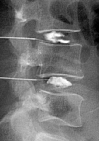
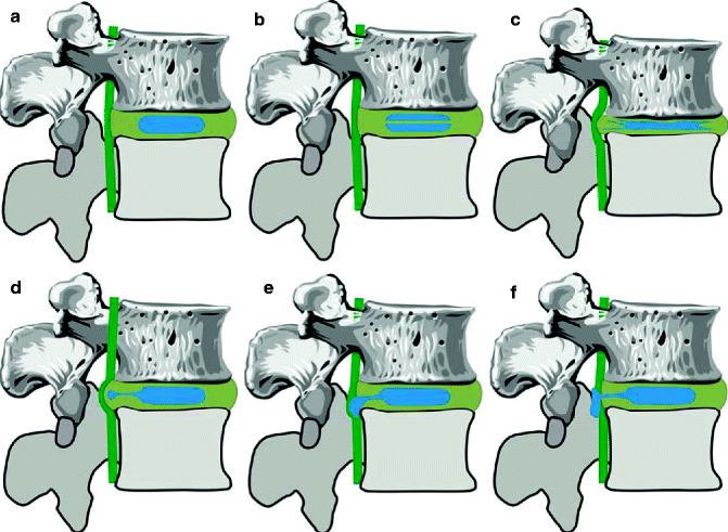
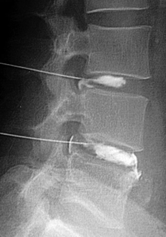
Fig. 41.6
Appearance of injected contrast agent in normal L3/4 (bilocular or hamburger bun) and L4/5 (unilocular or rectangular) discs
Fig. 41.7
Normal and abnormal patterns of disc morphology on the lateral radiographic projection. (a) Normal unilocular disc. (b) Normal bilocular disc. (c) Bulging disc with irregular annular fissures and loss of height. (d) Protruded disc with radial tear. (e) Extruded disc with inferior migration giving a “candle drip” appearance. (f) Extruded disc with disruption of the posterior longitudinal ligament (Reproduced with permission from Peh (2009))
Fig. 41.8
Radiographic appearance of abnormal discs on the lateral radiographic projection. The L3/4 disc has an annular tear with posterior protrusion. The L4/5 disc is degenerated with multiple anterior and posterior annular fissures, posterior protrusion, and extra-annular leakage
The Dallas discogram description is based on CT appearances and was originally classified into grades 0–3 (Sachs et al. 1987) and subsequently modified to four grades (Aprill and Bogduk 1992). Further modifications of the Dallas description included additional grades from 5 to 7 (Figs. 41.9 and 41.10).
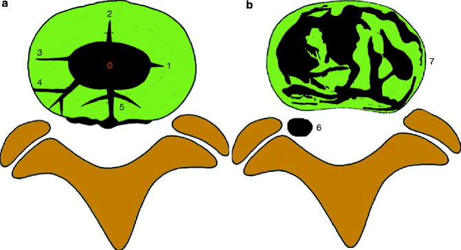
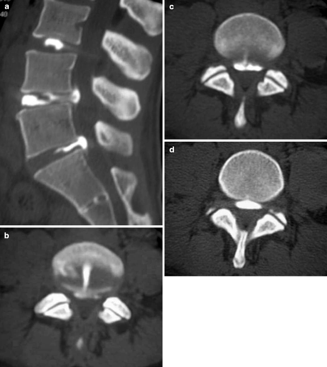
Fig. 41.9
Diagrammatic representation of the modified Dallas discogram description. (a) Grade 0 normal. Grade 1 fissure up to inner third. Grade 2 fissure up to middle third. Grade 3 fissure to outer third <30°. Grade 4 fissure to outer third >30°. Grade 5 full-thickness annular tear with extra-annular contrast extravasation. (b) Grade 6 disc sequestration and Grade 7 diffuse tears in degenerate disc
Fig. 41.10
CT lumbar discography. (a) Sagittal discogram images show a normal L3/4 disc (grade 0), a degenerate L4/5 disc with a posterior annular tear and posterior extrusion, and a L5/S1 posterior annular tear with posterior extrusion. (b–d) Axial discogram images of the L3/4 disc show a fissure extending through the posterior annulus of <30° circumference, with a resultant extra-annular contrast leakage posteriorly (extrusion) (grade 5)
Grade 0: Contrast agent is confined entirely within the normal nucleus pulposus.
Grade 1: Contrast agent extends radially along fissure involving the inner one-third of the annulus fibrosis.
Grade 2: Contrast agent extends into the middle one-third of the annulus fibrosis.
Grade 3: Contrast agent extends into the outer one-third of the annulus fibrosis, either focally or radially, to an extent not greater than 30º of the disc circumference.
Grade 4: Contrast agent extends into the outer one-third of the annulus fibrosis, dissecting radially to involve more than 30º of the disc circumference.
Grade 5: Represents a full-thickness tear, either focal or circumferential, with extra-annular contrast leakage (Schellhas et al. 1996).
Grade 6: Represents disc sequestration.
Grade 7: Represents a diffuse annular tear in disc degeneration (Bernard 1990).
Related posts:

Stay updated, free articles. Join our Telegram channel

Full access? Get Clinical Tree


