Fig. 42.1
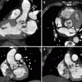
Biopsy devices. (a) BiopinceTM biopsy gun which obtains a 16-gauge sample by firing a hollowed cannula to obtain a core of tissue. (b) Temno® needle (18 gauge shown) with the inner stylet deployed. The needle’s outer cannula moves over the inner tray to obtain the sample. (c) Close-up of the inner stylet which will house the cut sample. (d) Coaxial needle (17 gauge shown) which may be initially advanced into a lesion and serve as the outer bore for biopsy devices including the Temno® and smaller FNAB needles
Whenever multiple biopsies are required, as in the case of most focal lesion biopsies, a coaxial needle (Fig. 42.1d) is often used. The core sampling device as well as the smaller-bore FNAB needles may be advanced within the coaxial needle. Placing the coaxial needle first avoids having to repeatedly puncture the capsule of an organ and expedites the biopsy itself as the lesion does not need to be reimaged and retargeted each time. Once the position of the coaxial needle has been assessed and the expected distance beyond its tip through which the biopsy devices pass is confirmed to be within the lesion, samples may be obtained without further imaging. However, it is good practice to show deployment of the biopsy devices within the lesion. It is also important to gently torque the coaxial needle into different portions of the lesion to prevent resampling the same tract and thereby potentially limiting diagnostic yield.
The handling of biopsy samples may be specific to a given institution’s pathology laboratory; however, typically core biopsy samples may be placed in formalin or saline and fine-needle aspirants expressed onto slides and/or cytologic preparatory material.
42.4 Organ-Specific Biopsy Approach
The general techniques of percutaneous biopsy apply to all lesions regardless of their location; however, lesions in different organs warrant specific approaches and have unique potential complications. Some lesions are not organ based and may exist as deposits within the peritoneal cavity or within the subcutaneous tissues or muscles. In all cases, typically the most direct route that avoids transgression of vital structures is the optimal trajectory. Some of the more common biopsy sites are detailed in this section.
42.4.1 Liver Biopsy
Liver biopsies may be either non-focal or focal, the former to provide overall assessment of the organ and latter to assess a specific lesion or lesions for malignancy or infection.
Non-focal biopsies are typically performed in the setting of abnormal liver function tests including the assessment of liver transplant rejection and deposition diseases such as hemochromatosis or Wilson disease. Non-focal biopsies are typically performed under ultrasound guidance and, depending on the interventional radiologist’s preference and the anatomy of the liver, can be aimed toward the right or left lobe via either an intercostal, subcostal, or subxiphoid approach (Fig. 42.2). Depending on the gauge of needle used and the diagnostic considerations, single or multiple cores may be procured. When planning the approach, care must be taken to identify and avoid the gall bladder, large vessels within the liver, as well as the closely apposed heart, right hemidiaphragm, and lung. Focal liver biopsy to assess for malignancy may be performed using ultrasound or CT with access gained via a coaxial needle through which core samples and FNABs are obtained (Fig. 42.3).
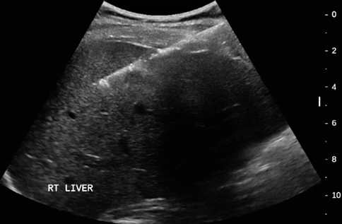
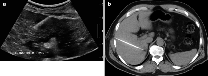
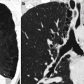
Fig. 42.2
Non-focal liver biopsy of the right lobe via an intercostal approach. Focal liver biopsies to assess for malignancy may be performed using ultrasound or CT with access via a coaxial needle through which core samples and FNABs are obtained (see also Fig. 42.3)
Fig. 42.3
Focal liver biopsies. (a) Ultrasound showing biopsy gun firing into a relatively peripheral hypoechoic liver lesion. (b) CT showing biopsy needle placement into a small hypoattenuating segment VII lesion that was not easily apparent on ultrasound. Image demonstrates deployment of the inner stylet of a side-cutting needle into the lesion
42.4.2 Renal Biopsy
Non-focal and focal biopsies of the kidney are commonly performed. Non-focal biopsies are often indicated in the case of renal failure with concern for intrinsic renal disease or to assess for rejection in the setting of renal transplant and typically involve obtaining core samples of renal cortex under ultrasound guidance with the patient in a prone position. The biopsy needle should be directed toward either the upper or lower pole, away from the renal hilum to avoid injury to the renal vasculature. Samples of a diameter of at least 16 gauge are usually obtained (Arellano 2011) (Fig. 42.4a).
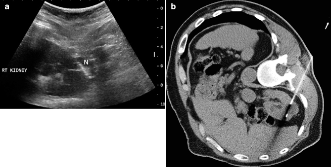
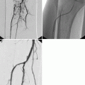
Fig. 42.4
Non-focal and focal renal biopsies. (a) Ultrasound-guided entry into the lower pole region of the right kidney with biopsy needle (N). (b) CT showing needle core biopsy of heterogeneous lower pole lesion
Focal renal biopsies are typically more amenable to CT-guided biopsy (Fig. 42.4b). Though patient positioning and trajectory may be dependent upon the location of the lesion within the kidney, placing the patient ipsilateral side down helps to stabilize the kidney and move the lower lobe of the lung out of the way.
Related posts:

Stay updated, free articles. Join our Telegram channel

Full access? Get Clinical Tree


