Fig. 27.1
MDCT coronal reconstruction of a 75-year-old man showing an appendiceal wall thickening, presence of an appendicolith, and signs of periappendiceal inflammation (arrow). At surgery: localized peritonitis secondary to acute perforated appendicitis
Fig. 27.2
MDCT coronal reconstruction showing a diffuse parietal thickening of some jejunal loops (white arrows), inflammatory changes of the mesenteric fat, and peritoneal fluid (black arrows). Diffuse peritonitis
27.2 Physiology of the Peritoneum
The abdominal cavity is covered by the parietal peritoneum, and it turns into the visceral peritoneum to cover the abdominal viscera. The total surface of the peritoneum is approximately 1.7 m2. In normal conditions it is sterile, and it contains 50 mL of yellow fluid, which contains a few macrophages, mesothelial cells generally, and lymphocytes. The initial response of the peritoneum against the bacterial contamination is characterized by hyperemia and increased exudates of fluid with phagocytes into the peritoneal cavity. In this initial stage these are predominately macrophages. Neutrophils arrive within 2–4 h and become the predominant cells of the peritoneal cavity by the first 48–72 h. These cells release great amounts of cytokines: the combined effect of these mediators contributes to observed inflammatory response during peritonitis. As a consequence of this inflammation, there is production of fibrinogen in the septic foci, with fast formation of fibrin, creating a mesh of fibrin that temporarily reduces and blocks the reabsorption of fluid from peritoneal cavity and “traps” bacteria. This phenomenon may generate the formation of an abscess. This response can be controlled and the peritonitis can be resolved, or it may continue to produce a residual or persistent peritonitis.
27.3 Classification
Peritonitis is classified as primary peritonitis and secondary peritonitis (Ordonez and Puyana 2006). Primary peritonitis is an inflammation of the peritoneum by an extraperitoneal source, frequently occurring from hematogen dissemination. It occurs in children and adults and can endanger life, particularly in patients who have cirrhosis or in children who have nephrosis. Secondary peritonitis is an infection that results from an inflammation or mechanical break of the integrity of the intestinal or the urogenital tract or solid organs, thus exposing the peritoneal cavity to the resident flora of the gastrointestinal tract. Secondary peritonitis is classified as acute peritonitis by perforation, postoperative peritonitis, or posttraumatic peritonitis.
27.4 Pathophysiologic Intra-abdominal Changes in the Older Adult
Older adults, defined as those who are 65 years and older, are the fastest growing segment of the population in the United States and the highest emergency department users of any age group. Abdominal pain is the most common presenting complaint to the emergency department, and in older adults, abdominal pain is the fourth most common chief complaint (Ragsdale and Southerland 2011). Pathophysiologic changes secondary to aging cause an increased susceptibility to intra-abdominal diseases as well as atypical clinical presentations. These changes occur from cellular to systemic levels, especially in the immune, genitourinary, gastrointestinal, nervous, and cardiovascular systems. The effects of aging on the GI system also predispose patients to abdominal pathologic conditions. The stomach has a slightly decreased emptying time. Acid secretion may increase secondary to decreased prostaglandin production. Liver mass and liver blood flow decrease with aging, resulting in decreased albumin synthesis. In the colon, the number of diverticula in the bowel increases with age. Because of physiologic anorexia of aging, there is decreased fluid and nutrient intake, predisposing the older adult to constipation.
27.5 Diagnosis
The diagnosis of peritonitis is a clinical diagnosis, based mostly on history and physical examination. The main symptom in all cases is abdominal pain. Tachycardia and the decrease in the amplitude of the pulse are indicatives of hypovolemia, and they are common in the majority of patients. Pain on palpation is the most characteristic sign of peritonitis, both deep and superficial touch. Bowel sounds may or may not be present, and they may resemble an early ileus. Localized peritonitis generates pain localized toward the inciting organ. Percussion of the abdomen may aid in accurately localizing the place of maximum peritoneal irritation. The patient may have an elevated white cell count greater than 11,000 cells per mL. Leukopenia suggests generalized sepsis and is associated with a poor prognosis. Blood chemistry may be normal, but in serious cases it may indicate severe dehydration, such as increased blood urea nitrogen and hypernatremia. Metabolic acidosis helps in the confirmation of the diagnosis. Urinalysis is indispensable to rule out urinary tract infection, pyelonephritis, and nephrolithiasis.
27.6 Challenges to Diagnosis in the Older Adult
Several variables create complexities in securing a diagnosis in the elderly patients. These include the physiologic changes that accompany aging, difficulties with taking an adequate history, medications that cause or confound pathology, lack of expected vital sign changes and physical findings, significant comorbidities, and seemingly normal laboratory values in the face of surgical disease. Acute abdomen in elderly patients poses a difficult challenge for emergency physicians. Elderly patients have a diminished sensorium, allowing pathology to advance to a very dangerous state before developing symptoms. In the presence of serious intra-abdominal pathology, elderly patients are more likely to present with vague symptoms and to have nonspecific findings on examination. Mesenteric ischemia and small bowel obstruction must be included in the initial differential diagnosis of abdominal pain, because an early diagnosis minimizes the risk of an unfortunate outcome (Reginelli et al. 2008). Elderly patients are also more likely to present with complications of abdominal surgical conditions. They are three times more likely to have a perforated appendix at surgery than the general population. Likewise, complications of acute cholecystitis are far more common in the elderly. Morrow et al. (1978) found that 40 % of acutely ill elderly patients who had cholecystitis had concomitant empyema of the gallbladder, gangrenous cholecystitis, free perforation, or subphrenic or hepatic abscess. The mortality of perforation in the general population is approximately 10 %, whereas in the geriatric population it is 30 % and increases eightfold if the diagnosis is delayed by 24 h due to peritonitis (Pinto et al. 2000; Grassi et al. 2004a). The type and degree of peritoneal contamination depends on the site, size, and duration of the perforation and on the physiologic state, including the time from the last meal, administration of a mechanical bowel preparation before the perforation, coexistent diseases, and the presence or absence of an ileus or bowel obstruction with accompanying bacterial overgrowth (Putcha and Burdick 2003; Guarner 2005). These factors affect the relative degree and type of bacterial and fungal contamination from perforation. The anatomic site of perforation significantly affects the type and burden of enteric contamination (Guarner 2005; Guarner and Malageleda 2003). The degree and type of microbiologic colonization of the gastrointestinal tract varies for different gastrointestinal organs and depends upon the local microenvironment. Microbiological colonization increases from proximal to distal, with the stomach showing the lowest number of viable microorganisms per cubic centimeter of luminal contents.
27.7 Imaging Techniques
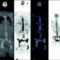
Radiological imaging is an important part of patient workup, separating the mundane causes from those requiring active medical intervention. In elderly patients with acute abdominal pain, the general usefulness of plain radiographs is limited mainly to evaluation for free intraperitoneal air, signs of obstruction, or foreign body ingestion or insertion. Although neither cost-effective nor diagnostically helpful as a general screening tool in the elderly patient with abdominal pain, there are several clues to serious disease that may be found on plain radiographs (Hendrickson and Naparst 2003): suggestion of cecal or sigmoid volvulus, signs of biliary tract disease such as emphysematous cholecystitis, and a calcified aneurysmal aorta. Free intraperitoneal air on plain radiographs is absent in 40 % of patients who have perforation (Pinto et al. 2000). When it is present it is often best visualized on supine decubitus films. Conventional radiography has been nowadays replaced by ultrasound, MDCT, and sometimes MR, in the evaluation of acute abdomen in the elderly patients. Ultrasound is the primary imaging modality of choice for the evaluation of right upper quadrant pain. Over the years, MDCT has gained a key role in the acute abdomen diagnostic process and especially in acute diseases of the small and large intestine. It has thus reduced the use of conventional radiology (Grassi et al. 2004b) due to its clear benefits of fast execution, broad view, and objective interpretation, as well as its ability to enable differential diagnosis in the event that the initial clinical suspicion is not confirmed especially in the elderly patients (Leschka et al. 2005). In elderly patients with diffuse peritonitis, the MDCT findings consist of thickening of the peritoneum and mesentery, increased density of the mesenteric fat, and ascites (Fig. 27.3


Related posts:
Stay updated, free articles. Join our Telegram channel
Full access? Get Clinical Tree
