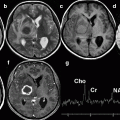
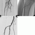
Fig. 8.1
Bone lesions at the Rizzoli Institute in patients over age 65 (Istituto Ortopedico Rizzoli – Laboratory of Experimental Oncology – Section of Epidemiology – Bologna, Italy)
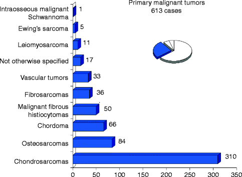
Fig. 8.2
Primary malignant bone tumors in patients over age 65 (Istituto Ortopedico Rizzoli – Laboratory of Experimental Oncology – Section of Epidemiology – Bologna, Italy)
8.2.1 Diagnosis
Analysis of bone tumors is statistical, leading to a reasonable probability of a correct diagnosis (Lodwick et al. 1980; Madewell et al. 1981). The radiologist and the pathologist both do the same job but with different tools. The radiologist studies the whole specimen from afar and the pathologist a small piece from close-up. Combining the two approaches brings the best results, emphasizing the importance of teamwork in providing the best care.
Age is very useful: after 65, the great majority of bone tumors are metastases or myeloma. Diagnosing metastases requires a perfect understanding of how they could change the treatment: in other words if no change in treatment is planned, they should not be looked for, unless they are painful. Among primary tumors, chondrosarcomas are the most frequent. Location helps: adamantinoma involves the anterior cortex of the shaft of the tibia, and in the epiphysis, giant cell tumor is a logical guess. Size is also helpful as small lesions are often benign. The lesion can be centered on the middle of the medullary cavity, often eccentric, rarely on the cortex or periosteum.
The matrix can be lytic, calcified, or ossified. Popcorn calcifications indicate cartilage (with nodules calcified at the periphery or in between them). The shape of periosteal bone formations indicates the mode of growth of the tumor. When thick, they indicate slow growth, perpendicular to the main axis of the bone, almost always malignant. A Codman triangle indicates a longitudinal periosteal reaction broken by tumor progression in its center. Cortical destruction and soft tissue involvement indicate aggressiveness. A thin calcified periosteal formation limiting the lesion indicates slow growth, even if the cortex has disappeared, and is probably a benign lesion. Conversely, tumor on both sides of a not yet destroyed cortex is a good sign of aggressiveness (Glass et al. 1986).
The radiologist must then, usually on radiographs first, diagnose the “leave me alone lesions,” for which nothing more is needed. They are rare in geriatric patients (osteochondromas, hemangiomas, chondromas, fibrous dysplasia) and are discovered incidentally.
If the diagnosis is difficult, because the site is difficult to study, the next step is CT (Regent et al. 1986). It gives better analysis of short and flat bones. Small calcifications, density to characterize water or fat, limited cortical erosions, or soft tissue involvement is better detected. The cartilaginous cuff of an osteochondroma is easily measured (Brown et al. 1986). MR is less useful, visualizing fluid-fluid levels, or the high T2 signal of cartilaginous nodules, or peritumoral edema.
If imaging suggests malignancy, staging should be performed, if possible before biopsy. Biopsy should be performed by a well-trained physician. It is surgical, or radiological, and the tract must be chosen in collaboration with the surgeon. Congelation and tattooing of the tract must be planned when necessary.
8.2.2 Staging
8.2.2.1 Localization
Locating the tumor is done with help of MRI MR (Kenney et al. 1981; Aisen et al. 1986; Bloem et al. 1988; Bohndorf et al. 1986). Medullary extension, and so the level of resection, is analyzed. Skip lesions are detected, as well as the relation to vessels and nerves. The main limitation is the study of the joints: when a tumor abuts on the cartilage, it may be very difficult to detect minimal joint involvement that could change surgery. Radiation therapy fields are best chosen.
8.2.2.2 General Staging
Bone metastases are detected by bone scintigraphy, total-body MRI, or PET, and pulmonary metastases by CT (Schima et al. 1994; Vanel et al. 1984). All nodules are not malignant, and if CT sensitivity is rather good, specificity is quite poor, and treatment is rarely changed by the detection of pulmonary nodules without histological proof. PET is not sensitive to small pulmonary nodules.
8.2.3 Treatment Effectiveness and Follow-Up
Some tumors receive neoadjuvant chemotherapy, but this is rare in geriatric patients. Decrease of peritumoral edema and of early MR uptake after contrast injection is correlated with efficient treatment (Picci et al. 2001; Verstraete et al. 1994; De Baere et al. 1992; van der Woude et al. 1994). After surgery with non-paramagnetic prostheses or fixations (usually titanium), MR can still be performed with few artifacts.
8.2.4 Most Frequent Examples
8.2.4.1 Malignant
Chondrosarcoma (Fig. 8.3)
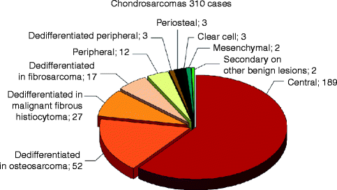
Fig. 8.3
Different types and frequency of chondrosarcomas in patients over age 65 (Istituto Ortopedico Rizzoli – Laboratory of Experimental Oncology – Section of Epidemiology – Bologna, Italy)
Conventional chondrosarcoma (Murphey et al. 1998; Geirnaerdt et al. 2000; Vanel et al. 2012) is a rather frequent tumor that can only be cured by surgery. Therefore, early diagnosis, clever biopsy tract, and perfect local staging are mandatory. On radiographs, the lesion is lytic, with typical round calcifications with a clear center, due to calcifications in between the lobules of cartilage or at the periphery of the lobules (Fig. 8.4). Limitation is usually sharp, indicating slow growth. Calcifications are better analyzed on CT, but a precise staging depends on MR. The tumor has a typical high signal on T2-weighted sequences, with no or limited uptake after injection, only around the lobules. Nodules of cartilage separated by fat marrow are often visible at the periphery of the main tumor, confirming the cartilaginous nature of the lesion, and correspond to a benign probably preexisting chondroma. Dedifferentiated chondrosarcoma (Staals et al. 2006, 2007) is revealed radiologically by a completely lucent area, without calcifications. On MR, this aggressive part takes up contrast medium, which helps guide the biopsy. Diagnosing the dedifferentiated part of the tumor is a useful part of the radiological work. Peripheral chondrosarcoma is often diagnosed on new pain or radiological changes of a known osteochondroma. The thick cartilaginous cuff, easily measured on ultrasound, CT, or MR, indicates malignancy. Dedifferentiation is possible but rare (Fig. 8.5).
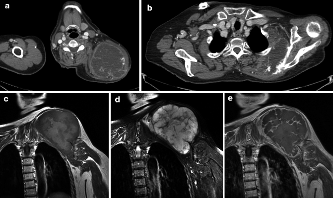
Fig. 8.4
Chondrosarcoma of the scapula. Injected CT (a, b), MRI coronal T1-weighted (c), and T2 fat-saturated (d) and injected (e) images. The tumor has a lobular pattern, hypodense on CT. The lesion starts from the spine of the scapula. It has a high signal and a lobular pattern on T2 images. Only the septae enhance after injection
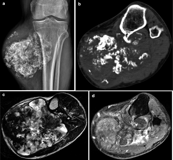
Fig. 8.5
Dedifferentiated peripheral chondrosarcoma of the tibia. On radiograph (a) and CT (b), typical cartilaginous calcifications, round with a clear center, are well seen. The basis of the osteochondroma is visible on CT. On an axial T2-weighted MRI (c), a part of the lesion is made of high-signal lobules, but the lesion is heterogeneous. The dedifferentiated part takes up strongly contrast medium after injection (d). The biopsy must be directed at this part
Osteosarcoma (Fig. 8.6) can be conventional or often secondary to a preexisting lesion, infarct, Paget disease (Shaylor et al. 1999) (Fig. 8.7), or radiation changes (Fig. 8.8). Soft tissue involvement and cortical lysis indicate fast growth. Fat disappearance is a reliable indicator of malignant changes. Cartilage inside a fibrous dysplasia usually does not indicate malignant changes but only a cartilaginous benign component inside the lesion.
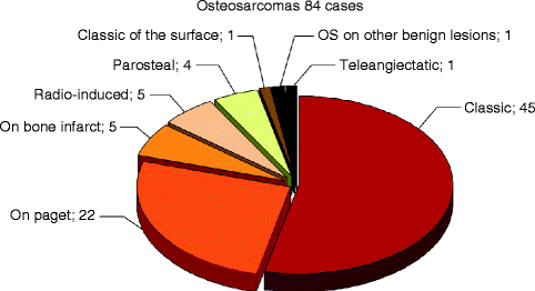
Fig. 8.6
Different types and frequency of osteosarcomas in patients over age 65 (Istituto Ortopedico Rizzoli – Laboratory of Experimental Oncology – Section of Epidemiology – Bologna, Italy)
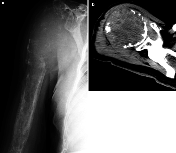
Fig. 8.7
Osteosarcoma on Paget disease. On radiograph (a), cortical thickening typical of Paget disease is easily recognized. The proximal humerus is completely lytic. On CT (b), the tumor is heterogeneous, with fluid-fluid levels, and corresponds to a telangiectatic osteosarcoma
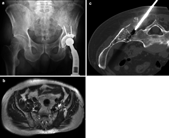
Fig. 8.8
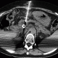
Radiation-induced osteosarcoma after treatment of a bone lymphoma. On radiograph (a), there is a hip prosthesis following radiation-induced osteonecrosis. On axial T1-weighted MRI, a part of the fat marrow is replaced (b). The CT-guided biopsy (c) confirmed an osteosarcoma
Fibrosarcoma is an aggressive, purely lytic, often metadiaphyseal tumor. It can also develop on an infarct.
Chordoma involves the sacrum, clivus, and rarely mobile spine. Surgery is the only curative treatment. It is difficult in old patients. The lesion is lytic and often contains remaining pieces of bone. MR is the key examination for local staging, allowing à reliable prediction of the hazards and probable sequels (Fig. 8.9). Metastases are rare.
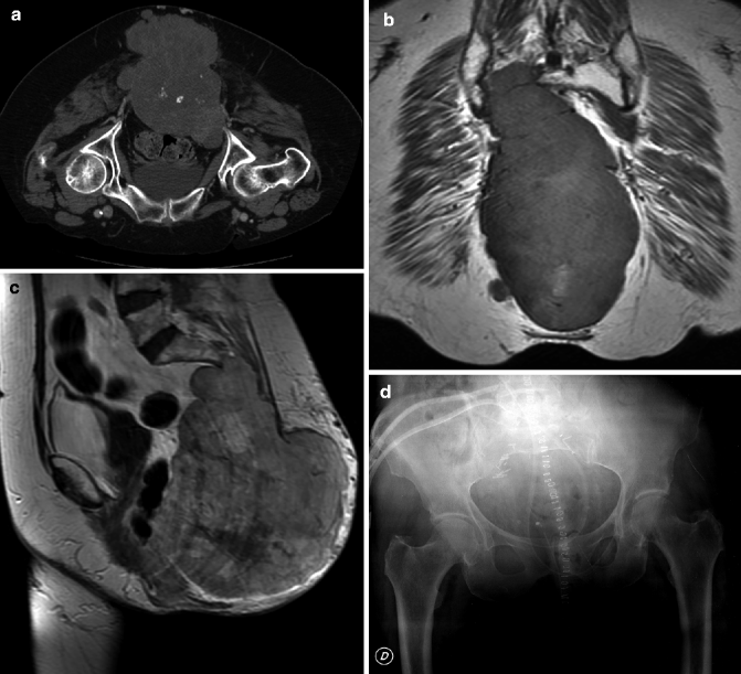
Fig. 8.9
Chordoma. On CT (a), the huge mass contains remaining pieces of bone. Coronal T1-weighted (b) and sagittal injected (c) images analyze the extension and allow a choice of level of resection. On the postoperative radiograph (d), S1 is preserved
Plasmocytoma is rare over 65 (Fig. 8.10). It is a purely lytic well-limited lesion, containing often thick trabeculae. Whole-body MR allows detection of minimal bone marrow lesions in about half of the cases, allowing a general treatment. Myeloma (Fig. 8.11) has multiple, purely lytic, well-limited lesions. Evaluation of the tumor mass is still performed on radiographs, even if MR detects more lesions. Imaging helps follow the treatment and detect complications (Baur et al. 2002a; Lecouvet 2001).
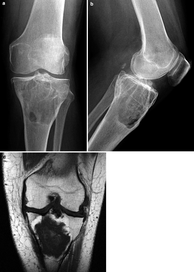
Fig. 8.10
Plasmocytoma. On radiographs (a AP and b lateral), a lytic well-limited lesion with thick trabeculae is well analyzed. It is central and has a low signal on T1-weighted coronal MRI (c)
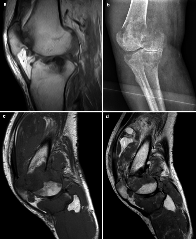
Fig. 8.11
Myeloma. Sagittal T1-weighted images (a), showing minimal femoral and tibial lesions, considered as traumatic. Three months later, fracture and lytic tibial and femoral lesions, on radiograph (b), sagittal T1 (c), and T2-weighted (d) images
8.2.4.2 Benign Tumors
They are incidental findings or are known for a long time. An asymptomatic chondroma is frequently discovered incidentally on MR performed for another reason in the knee or shoulder of a patient. A lesion centered in the medullary cavity without or with a limited cortical involvement and no soft tissue extension is very probably benign and does not require further examination.
Giant cell tumors can be found in the metaphyses of long bones and pelvis. They are purely lytic, well limited but without sclerosis. Denosumab may change their treatment. They may rarely develop in Paget disease.
8.3 Metastases
8.3.1 Introduction
They are by far the most frequent bone tumors in old patients. Imaging should be used for routine screening in tumors with a high incidence of bone metastases such as breast and prostate cancer, if and only if there are practical consequences (Boccardo et al. 1995; Rosselli Del Turco et al. 1994). It is used in symptomatic patients presenting with pain or neurological deficit, and it is mandatory before aggressive surgery (pneumonectomy, prostatectomy, cystectomy, amputation, etc.), when their discovery may change the surgical approach. For these purposes, imaging includes radiographs, radionuclide scintigraphy, CT, MRI, and PET.
Most bone metastases are asymptomatic. Over 90 % occur within the red marrow (Silverberg and Lubera 1987; Abrams et al. 1950; Krishnamurthy 1977). That explains why systematic detection looks mainly at the spine, pelvis, and proximal long bones. Approximately 10 % of metastases are solitary, which may rarely have practical treatment consequences: an isolated bone metastasis can be cured in 40 % of kidney cancers, but not in a breast cancer. Extraskeletal spread into the soft tissues is rare (Wyche and De Santos 1978). Special imaging features can guide the search of a primary unknown cancer: distal metastases (Fragiadakis and Panayotopoulous 1972) and cortical bone metastases most frequently occur in lung cancer (Deutsch and Resnick 1980


Stay updated, free articles. Join our Telegram channel

Full access? Get Clinical Tree


