, Joungho Han2, Man Pyo Chung3 and Yeon Joo Jeong4
(1)
Department of Radiology Samsung Medical Center, Sungkyunkwan University School of Medicine, Seoul, Korea, Republic of (South Korea)
(2)
Department of Pathology Samsung Medical Center, Sungkyunkwan University School of Medicine, Seoul, Korea, Republic of (South Korea)
(3)
Department of Medicine Division of Pulmonary and Critical Care Samsung Medical Center, Sungkyunkwan University School of Medicine, Seoul, Korea, Republic of (South Korea)
(4)
Department of Radiology, Pusan National University Hospital, Busan, Korea, Republic of (South Korea)
Abstract
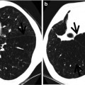
An air crescent is a collection of air in a crescentic shape that separates the wall of a cavity from an inner mass [1, 2] (Fig. 14.1).
Definition
An air crescent is a collection of air in a crescentic shape that separates the wall of a cavity from an inner mass [1, 2] (Fig. 14.1).
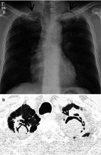
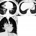
Fig. 14.1
Air-crescent sign in aspergillomas growing within chronic tuberculous cavities in a 76-year-old man. (a) Chest radiograph shows air-crescent signs (arrows) in both apices. Also note small nodules in the bilateral upper lung zones. (b) Lung window image of thin-section (1.5-mm section thickness) CT scan obtained at level of great vessels demonstrates aspergillomas within cavities disclosing so-called air-crescent signs (arrows) in both lung apices. Also note parenchymal band in the right apex and bullae in both apices
Diseases Causing the Sign
The air-crescent sign is often considered characteristic of either Aspergillus colonization of preexisting cavities (aspergilloma) (Figs. 14.1 and 14.2) or retraction of infracted lung in angioinvasive pulmonary aspergillosis. Other less common causes include tuberculosis, Rasmussen’s aneurysm (Fig. 14.3), ANCA-associated granulomatous vasculitis (former Wegner’s granulomatosis), complicated hydatid disease, hematoma, lung abscess, and necrotic lung cancer (Table 14.1).
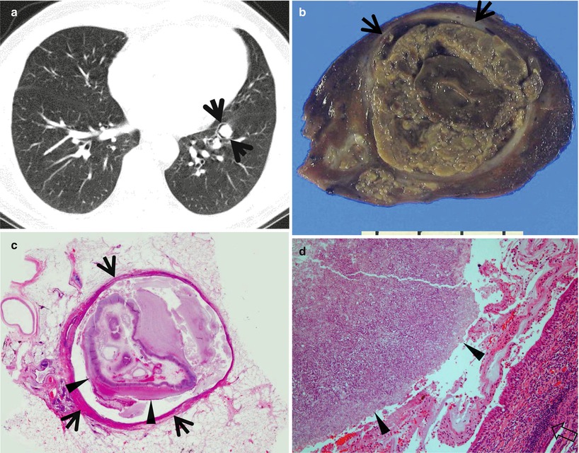
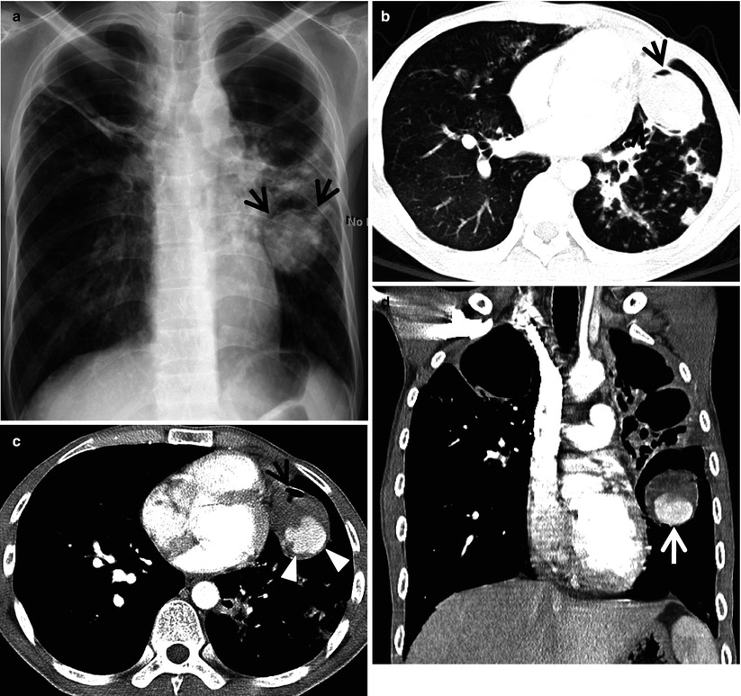
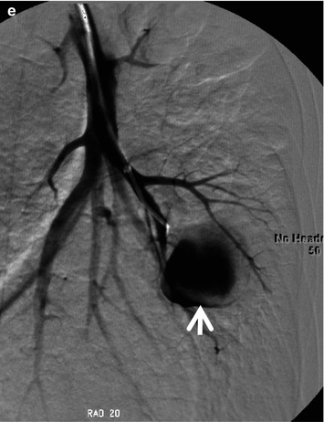
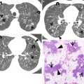
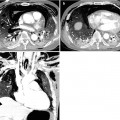
Fig. 14.2
Air-crescent sign in an aspergilloma growing within a dilated bronchus in a 53-year-old man. (a) Lung window image of thin-section (1.5-mm section thickness) CT scan obtained at level of right inferior pulmonary vein shows an aspergilloma showing air-crescent sign within a dilated bronchus (arrows) in left lower lobe. (b) Gross pathology of resected specimen (left lower lobectomy) demonstrates a soft yellow-gray lesion (arrows) within a dilated bronchus. Air is located between the lesion and airway wall, and the air is the source of air-crescent sign on CT scan. (c) Scanning view exhibits a dilated bronchus (arrows) harboring a fungus ball (arrowheads). (d) High-magnification (×100) photomicrograph discloses a part of aspergilloma consisting of dense clump of fungal hyphae (arrowheads). Bronchial wall shows chronic inflammation (open arrow)
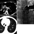
Fig. 14.3
Air-crescent sign in a Rasmussen’s aneurysm in a 34-year-old man. (a) Chest radiograph shows destructive tuberculous lesions in the bilateral upper lung zones in which bullae are also seen. Also note a mass showing air-crescent sign (arrows). (b) Lung window image of thin-section (2.5-mm section thickness) CT scan obtained at level of right inferior pulmonary vein shows a soft tissue mass depicting air-crescent sign (arrows) in lingular division of left upper lobe. Also note tuberculous lesions appearing as cavitating and noncavitating nodules in the left lung and right middle lobe. (c) Mediastinal window image of enhanced CT scan obtained at same level to (a) demonstrates a highly enhancing lesion of aneurysm (arrowheads) with surrounding air crescent (arrow). (d) Coronal reformatted image (2.0-mm section thickness) exhibits the aneurysm (arrow). (e) Left pulmonary angiogram discloses the aneurysm (arrow) more clearly
Table 14.1
Common diseases manifesting as air-crescent sign
Disease | Key points for differential diagnosis |
|---|---|
Aspergilloma | Intracavitary non-enhancing soft tissue mass withair-crescent sign |
Angioinvasive pulmonary aspergillosis | Retraction of infracted lung, CT halo sign |
Tuberculosis | |
Rasmussen’s aneurysm | Focal dilatation of one of the pulmonary segmentalarteries adjacent to tuberculous cavity |
ANCA-associated granulomatous vasculitis | Multiple, bilateral, subpleural nodules, or masses |
Complicated hydatid disease | Water-lily sign, homogeneous water density |
Hematoma | |
Lung abscess | |
Necrotic lung cancer |
Distribution
Related posts:
Stay updated, free articles. Join our Telegram channel

Full access? Get Clinical Tree


