, Joungho Han2, Man Pyo Chung3 and Yeon Joo Jeong4
(1)
Department of Radiology Samsung Medical Center, Sungkyunkwan University School of Medicine, Seoul, Korea, Republic of (South Korea)
(2)
Department of Pathology Samsung Medical Center, Sungkyunkwan University School of Medicine, Seoul, Korea, Republic of (South Korea)
(3)
Department of Medicine Division of Pulmonary and Critical Care Samsung Medical Center, Sungkyunkwan University School of Medicine, Seoul, Korea, Republic of (South Korea)
(4)
Department of Radiology, Pusan National University Hospital, Busan, Korea, Republic of (South Korea)
Abstract
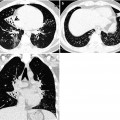
The signet ring sign is a combination resembling a signet (or pearl) ring, which is composed of a ring-shaped opacity representing a dilated bronchus in cross section and a smaller adjacent opacity representing its pulmonary artery [1] (Fig. 15.1). Normally, the diameter of a bronchus is equal to the diameter of the adjacent pulmonary artery. The signet ring sign occurs when the bronchoarterial ratio is increased. It is the basic CT sign of bronchiectasis.
Definition
The signet ring sign is a combination resembling a signet (or pearl) ring, which is composed of a ring-shaped opacity representing a dilated bronchus in cross section and a smaller adjacent opacity representing its pulmonary artery [1] (Fig. 15.1). Normally, the diameter of a bronchus is equal to the diameter of the adjacent pulmonary artery. The signet ring sign occurs when the bronchoarterial ratio is increased. It is the basic CT sign of bronchiectasis.
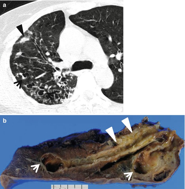
Fig. 15.1
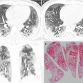
Bronchiectasis showing signet ring sign in a 44-year-old man. (a) Lung window image of CT scan (2.5-mm section thickness) obtained at level of the right upper lobar bronchus shows dilated bronchi showing signet ring sign (arrows) in right upper lobe. Also note mucus plugging (arrowhead) in dilated bronchi. (b) Gross pathologic specimen obtained with right upper lobectomy discloses cylindrical bronchiectasis and distal cystic changes (arrows, cystic bronchiectasis). Also note thickened bronchial wall (arrowheads) with active inflammation
Diseases Causing the Sign
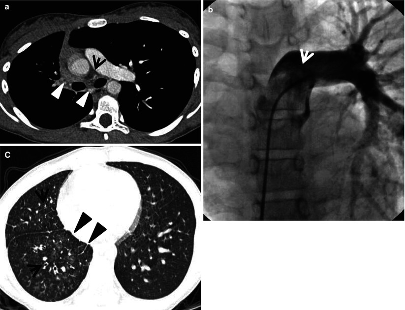
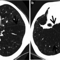
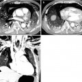
Although the signet ring sign is the basic CT sign of bronchiectasis, it can also be seen in diseases characterized by abnormal reduced pulmonary arterial flow (e.g., proximal interruption of pulmonary artery or chronic thromboembolism) (Fig. 15.2) (Table 15.1).
Get Clinical Tree app for offline access
Fig. 15.2
Proximal interruption of pulmonary artery showing signet ring sign in a 10-year-old girl. (a) Mediastinal window image of enhanced CT scan (5.0-mm section thickness) obtained at level of the main bronchi shows interruption of right proximal pulmonary artery (arrow) within pericardium. Also note hypertrophied right bronchial artery and branches (arrowheads). (b) Conventional pulmonary angiograph demonstrates interrupted right proximal pulmonary artery (arrow). (c) Lung window image obtained at ventricular level displays signet ring sign (arrows) constituted by dilated bronchus and accompanying small systemic artery in the right lung. Also note smooth interlobular septal thickening (arrowheads


Related posts:
Stay updated, free articles. Join our Telegram channel
Full access? Get Clinical Tree
