Airway


Airway


Evan J. Zucker

Supika Kritsaneepaiboon

Omolola M. Atalabi

Ricardo Restrepo

Yumin Zhong

Sally A. Vogel

Edward Y. Lee


INTRODUCTION

Airway disease is common in the pediatric population, and imaging is an essential component of the diagnostic evaluation. Prompt recognition and accurate diagnosis are crucial, as many disorders can be potentially life threatening. Affected pediatric patients often present with stridor, wheezing, and respiratory distress because of acute airway obstruction. With smaller and more collapsible airways, infants and children tend to develop symptoms earlier than do their adult counterparts.1 Chronic airway obstruction may manifest as recurrent pulmonary infections or obstructive sleep apnea (OSA).2

In this chapter, an overview of pediatric airway disease is provided. First, the variety of imaging techniques currently available for evaluating the pediatric airway is discussed. The normal anatomy of the pediatric airways is reviewed. Finally, selected pediatric airway disorders important for everyday practice, focusing on pathophysiology, clinical features, imaging assessment, and treatment approaches, are presented.


IMAGING TECHNIQUES


Radiography

In pediatric patients, who present with potential airway disorder, imaging assessment typically begins with frontal and lateral views of the neck and/or chest (Figs. 8.1, 8.2, 8.3, 8.4 and 8.5). Relatively inexpensive and widely available radiographs are the first-line modality for helping to exclude foreign body aspiration and other causes of respiratory distress.1,2

The airway is best evaluated with a magnification highkilovoltage (kV) technique with the anteroposterior (AP) view tightly coned to the neck and selectively filtered to eliminate overlying bony shadows from the cervical spine3 (Table 8.1). Proper positioning is essential for the lateral neck view, which should be performed in inspiration with moderate neck extension. Rotation, flexion, or expiration may lead to inaccurate interpretation1,2 (Fig. 8.4). However, an optimal exam may be difficult to achieve in
actual practice in the moving, crying infant. To help solve this problem, a variety of immobilization devices are available for children <4 years old. Pacifiers are also useful. An important caveat is that pediatric patients with suspected airway obstruction should never be forced into a position they do not wish to assume, because this may lead to acute respiratory decompensation, which is potentially life threatening.


FIGURE 8.1 Position for lateral airway radiography in an infant. The lateral radiograph is obtained in deep inspiration with the neck extended. Radiographer can stabilize the optimal neck position of the patient while obtaining the neck radiograph.


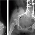
FIGURE 8.2 Lateral and anteroposterior airway radiographs. A: Lateral radiograph shows normal degree of oropharyngeal distension, “small finger-sized” epiglottis (curved arrow) and thin aryepiglottic folds (straight arrow). Moderate enlargement of adenoids (asterisk) is also seen. B: Anteroposterior airway radiograph demonstrates normal symmetric subglottic “shoulders” (arrows).

Chest radiographs should be obtained near the end of quiet inspiration. In infants, supine AP views of the chest are more easily obtained and adequate because the degree of magnification is similar compared to AP or posteroanterior (PA) erect radiographs. Lateral radiographs are rarely needed. In cooperative children (generally >4 years of age), PA and lateral erect views should be obtained, either standing or sitting (Figs. 8.5 and 8.6). Evaluation for air trapping, an indirect sign of tracheobronchial foreign body obstruction, can be further evaluated with expiratory views in cooperative older children (Fig. 8.7) or the less technically challenging lateral decubitus views in younger uncooperative patients1 (Fig. 8.8). Gonadal shielding is used to help limit radiation exposure.


FIGURE 8.3 Well-coned anteroposterior chest radiograph. Radiographer can stabilize the child for the optimal chest position while obtaining chest radiograph. Note the lead drape over the lower abdomen.


Ultrasound

Although in general an ideal pediatric imaging modality due to the lack of ionizing radiation, no need for sedation, and ability for real-time assessment in multiple planes, ultrasound (US) has traditionally played a limited role in evaluating the airway. Ultrasound is useful for assessing neck masses, helping to distinguish cystic from solid masses. Lesion vascularity and the patency of vessels and vascular catheters can be readily assessed with Doppler ultrasound.

Although not widely used, techniques for sonographic airway assessment have been described. Patients are imaged supine with a pillow under the shoulders, the head extended, and the neck flexed (“sniffing” position). A linear high-frequency transducer is best for superficial airway structures (within 2 to 3 cm from the skin), with images obtained in the transverse plane (Fig. 8.9). A curved low-frequency transducer is most useful for sagittal and parasagittal views of structures in the submandibular and supraglottic regions. Sonography complements the physical examination and direct visualization methods such as laryngoscopy. Ultrasound may also provide imaging guidance for procedures such as percutaneous tracheostomy.4,5


FIGURE 8.4 Suboptimal quality lateral airway radiograph due to overlying earrings and fingers with suboptimal neck extension obtained in expiration. Widening (asterisk) of the retropharyngeal soft tissues during expiration may mimic a retropharyngeal abscess.


Airway Fluoroscopy

Airway fluoroscopy is useful for evaluating dynamic abnormalities such as laryngomalacia and tracheomalacia (Fig. 8.10). It may be combined with a barium swallow study.2 The entire airway from nasopharynx to main bronchi can be evaluated safely, rapidly, and noninvasively in frontal, lateral, and oblique projections. In accordance with the As Low As Reasonably Achievable (ALARA) principle, radiation dose reduction techniques should be implemented whenever possible such as pulsed fluoroscopy and restricting time spent using the fluoroscopic pedal. Evaluation may be limited in uncooperative infants and young children and older children with larger body habitus.1,2


TABLE 8.1 Radiography Technique for Soft Tissue Neck


Age


View


KVP


MAS


AEC


Grid


SID


FSS


0-3 mo


AP


60


1.5


Off


No


40″


Small


3-12 mo


AP


60


2


Off


No


40″


Small


1-3 y


AP


65


2


Off


No


40″


Small


3-6 y


AP


65


5


Active


Grid


40″


Small


6-10 y


AP


70


5


Active


Grid


40″


Small


>10 y


AP


75


8


Active


Grid


40″


Small


Age


View


KVP


MAS


AEC


Grid


SID


FSS


0-3 mo


Lateral


60


4


Off


No


72″


Small


3-12 mo


Lateral


60


5


Off


No


72″


Small


1-3 y


Lateral


65


6


Off


No


72″


Small


3-6 y


Lateral


65


6.5


Off


No


72″


Small


6-10 y


Lateral


70


7


Off


No


72″


Small


>10 y


Lateral


75


15


Off


Grid


72″


Large


KVP, kilovoltage; MAS, current; AEC, automatic exposure control; SID, source to image-receptor distance; FSS, focal spot size.


Dynamic sleep airway fluoroscopy is useful for assessing such conditions as OSA. The patient is sedated with continuous vital sign and pulse oximetry monitoring. When signs of airway occlusion occur, fluoroscopic evaluation for 10 to 20 seconds is performed at selected anatomical sites in the lateral supine position and correlated with clinical findings. The oropharynx at the level of the base of the tongue, hypopharynx, and intrathoracic trachea is typically assessed. Downward pulling of the arms improves neck visualization, whereas an arms-above-head position maximizes assessment of the intrathoracic trachea. A maximum of 2-minute fluoroscopy is used to limit radiation exposure.6


Computed Tomography

Multidetector computed tomography (MDCT) with multiplanar two-dimensional (2D) and three-dimensional (3D) reformation is now the noninvasive gold standard for evaluation of the pediatric airway (Fig. 8.11). Combining precise anatomical detail with rapid scan times, MDCT often eliminates the need for sedation and intubation to achieve diagnostic imaging. Newer techniques including paired inspiratory-expiratory MDCT, cine MDCT, and four-dimensional (4D) MDCT permit dynamic airway assessment.7,8,9 Nevertheless, use of CT is tempered by growing concerns about risks of radiation exposure particularly in children.10


FIGURE 8.5 Positioning for upright chest radiograph in cooperative older children. Posteroanterior (A) and lateral (B) radiograph in an upright position can be obtained in sitting position and with the arm elevated.


FIGURE 8.6 Normal chest radiograph in an older child. A: Posteroanterior chest radiograph shows trachea (T), carina (C), and bilateral mainstem bronchi (B). B: Lateral chest radiograph demonstrates trachea (T) and carina (C).


FIGURE 8.7 Chest radiographs obtained at end inspiration and end expiration in a 5-year-old girl. This patient was subsequently diagnosed a nonradiopaque foreign body lodged in the left mainstem bronchus by bronchoscopy. A: Frontal chest radiograph obtained at end inspiration shows fairly symmetric lung aeration. B: Frontal chest radiograph obtained at end expiration demonstrates expected decreased right lung volume but substantial hyperinflation of the left lung indicating underling air trapping due to left mainstem bronchial obstruction.


FIGURE 8.8 Value of lateral decubitus view. A 2-year-old girl who presented with acute wheezing and coughing after playing with her brother’s plastic building toys. The patient was subsequently diagnosed with a nonradiopaque foreign body lodged in the left mainstem bronchus by bronchoscopy. A: Frontal radiograph shows mildly hyperinflated left lung compared to the right lung. B: Left-sided lateral decubitus view demonstrates persistent hyperinflation of the left lung.

Specific parameters for large airway imaging are dependent on the type of CT scanner. However, in general, optimal imaging can be achieved with ≥16-row MDCT and the following parameters: 0.75-mm collimation for 16-MDCT scanner, 0.625-mm collimation for 32-MDCT scanner, and 0.6-mm collimation for 64-MDCT scanner; high-speed mode; and a pitch equivalent of 1.0 to 1.5. Use of age- or weight-adjusted milliamperage (mA), lowest possible kV, and anatomically based real-time automated exposure control helps reduce radiation exposure. The intrinsic contrast between the air-filled large airways and adjacent mediastinal soft tissues also allows for lower-dose technique. Suggested parameters for tube current and kV from Boston Children’s Hospital are shown in Table 8.2. In addition to traditional coronal and sagittal reconstructions, curved planar reformats along the long axis of the trachea and bronchi can be routinely obtained to allow more accurate measurements of the trachea and bronchi1,2,7,11 (Fig. 8.11D).


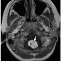
FIGURE 8.9 Normal trachea seen on ultrasound. Air within the nondependent portion of the trachea (T) in supine position of the patient shows increased echogenicity (arrow) with posterior shadowing. Also seen is normal thyroid gland (asterisks) on both sides of the trachea.


Magnetic Resonance Imaging

Magnetic resonance imaging (MRI) offers excellent contrast resolution without ionizing radiation. However, long scan times may require sedation to prevent patient motion. MRI of the neck and upper airway generally takes 30 to 60 minutes with current techniques (Fig. 8.12). Intravenous gadolinium contrast is useful for assessing suspected neoplastic or infectious causes of upper airway obstruction.2

Beyond static MRI for anatomical evaluation, cine MRI utilizing fast gradient-echo sequences is an emerging modality allowing dynamic airway assessment for such conditions as OSA and velopharyngeal insufficiency.2,12,13,14,15,16,17 Studies can be performed on either a 1.5 or 3.0 Tesla (T) MRI unit. The patient is sedated and placed in the head-and-neck vascular coil. In small pediatric patients, the airway from the superior nasal passages to the carina can be visualized in its entirety. In larger pediatric patients, the inferior aspect of the trachea may be outside the field of view. After a 3D localizer image is obtained, sagittal and axial T1-weighted spin-echo (SE) (representative parameters: repetition time [msec]/echo time [msec] of 400/minimal, 22-cm field of view, 4-mm section thickness with 1-mm gap, 256 × 192 matrix, two signals acquired) and sagittal and axial fast SE inversion recovery (IR)
(representative parameters: 5,000/34, echo train length of 12, 22-cm field of view, 6-mm section thickness with 2-mm gap, 256 × 192 matrix, two signals acquired) sequences are obtained. Cine MR is performed in midline sagittal and axial planes at the midportion of the tongue using a fast gradient-echo sequence (representative parameters: 8,200/3,600, 80-degree flip angle, 12-mm section thickness) with 128 consecutive images captured in ˜2 minutes and viewed on cine mode.2,16


FIGURE 8.10 Tracheomalacia in a 4-year-old girl who presented with chronic cough and recurrent pulmonary infection. Subsequently performed bronchoscopy confirmed the diagnosis of marked tracheomalacia. A: Lateral radiograph obtained at end inspiration during airway fluoroscopy study of airway shows patent trachea (arrows). B: Lateral radiograph obtained at end expiration during airway fluoroscopy study demonstrates marked (>75%) collapse of the trachea (arrows), consistent with tracheomalacia.


NORMAL ANATOMY AND VARIANTS

The airway includes the nose, paranasal sinuses, pharynx (nasopharynx, oropharynx, and hypopharynx), larynx, trachea, main bronchi, peripheral bronchi, and bronchioles. The nasopharynx is located posterior to the nasal cavity and superior to the soft palate (Fig. 8.12). The oropharynx is situated between the soft palate and tip of the epiglottis. The hypopharynx (laryngopharynx) is bounded by the tip of the epiglottis and the cricoid cartilage. The larynx, bounded by the cricoid cartilage and the tongue base, includes the thyroid and cricoid cartilage, paired arytenoids, and epiglottis.18 The supraglottic larynx includes the epiglottis, aryepiglottic folds, and false vocal cords, extending to the laryngeal ventricle. The glottic larynx extends from the laryngeal ventricle to the inferior margin of the true vocal cords. Finally, the subglottic larynx then extends to the level of the inferior margin of the cricoid cartilage, subglottic larynx, and upper trachea.

The trachea, a cartilaginous and membranous tube, extends from the larynx at approximately the level of C6 to the upper border of T5 (Fig. 8.13). There it divides into the right and left main bronchi. The right main bronchus divides into the right upper lobe bronchus and bronchus intermedius, which further branches into the right middle and lower lobe bronchi (Figs. 8.13 and 8.14). The left main bronchus divides into the left upper and lower lobe bronchi. There are three right upper lobe segmental bronchi (apical, anterior, and posterior), two right middle lobe segmental bronchi (lateral and medial), and five right lower lobe segmental bronchi (superior, medial basal, anterior basal, lateral basal, and posterior basal). There are four left upper lobe segmental bronchi (apicoposterior, anterior, superior lingular, and inferior lingular) and four left lower lobe segmental bronchi (superior, anteromedial, lateral basal, and posterior basal) (Figs. 8.13 and 8.14). Many normal variations in airway branching anatomy exist.

In initially assessing the upper airway, the lateral neck radiograph is most useful. On a normal exam, the following structures can be identified: nasopharynx, adenoids, hard

and soft palate, uvula, oropharynx, tongue, mandible, base of the tongue, vallecula, epiglottis, aryepiglottic folds, pyriform sinuses, laryngeal ventricle, true and false vocal cords, subglottic larynx, and upper trachea (Fig. 8.2).


FIGURE 8.11 Normal large airway in a 6-year-old girl. A: Enhanced axial CT image at the aortic arch level shows normal round and patent trachea (T) obtained at end inspiration. (A, aortic arch; SVC, superior vena cava; E, esophagus.) B: Enhanced axial CT image shows normal and patent bilateral mainstem bronchi (MB). (AA, ascending aorta; DA, descending aorta; SVC, superior vena cava; LP, left main pulmonary artery.) C: Sagittal reformatted lung window CT image of the large airway. A reference line (yellow line and red asterisks) through the center of the airway for reconstruction of a curved coronal reformatted CT image. D: Curved coronal reformatted CT image shows a straightened view of the entire trachea. E: 3D external volume-rendered CT image (i.e., virtual bronchography) of normal airway. (Continued)


FIGURE 8.11 (Continued) F: 3D internal volume-rendered CT image (i.e., virtual bronchoscopy) of airway obtained at glottis level. Mildly opened glottis is seen. G: 3D internal volume-rendered CT image of airway obtained at the level of carina. Bilateral mainstem bronchi are patent.

In children 5 years old or less, lateral deviation of the trachea is normal and should not be mistaken for pathology (Fig. 8.15). This phenomenon, which tends occur at or just above the thoracic inlet opposite the side of the aortic arch, is felt possibly because of the relatively long tracheal length with respect to the child’s short neck and rib cage.1,19 Other common normal variants are anterior buckling of the trachea and widening of the retropharyngeal soft tissues during expiration and neck flexion, features that may mimic a retropharyngeal abscess20 (Fig. 8.4).


TABLE 8.2 Tube Current and kV by Patient Weight for Central Airway MDCT


Weight (kg)


Tube Current (mAs) Insp./Exp.


kV


<10


40/20


80


10-14


50/25


80


15-24


60/30


80


25-34


70/35


80


35-44


80/40


80


45-54


90/40


90


55-70


100-120/40


100-120


For tube current and kilovoltage by patient weight for end-expiratory MDCT examination, mAs should be reduced by 50% to a maximum of 40 mA while maintaining the same level of kV for end-inspiratory MDCT examination.


Insp, inspiratory; Exp, expiratory; mA, milliamperage; kV, kilovoltage; MDCT, multidetector computed tomography.


Reprinted from Lee EY, Boiselle PM. Tracheobronchomalacia in infants and children: multidetector CT evaluation. Radiology. 2009;252:7-22, with permission.


SPECTRUM OF PEDIATRIC AIRWAY DISORDERS


Congenital and Developmental Anomalies


Choanal Atresia/Stenosis

Choanal atresia is a congenital obstruction of the nasopharynx characterized by narrowing and closure of the posterior choanae and medialization of the pterygoid plates and lateral wall of the nose.2,21 It is the most common etiology of neonatal nasal obstruction with an estimated incidence of 1 in 5,000 to 1 in 9,000 births, occurring twice as often in females as in males, more often unilateral than bilateral.2,21,22 Although osseous obstruction of the choanae was previously thought most common (90%) (Fig. 8.16), with membranous obstruction in the remaining 10% (Fig. 8.17), more recent data suggest a mixed bony/membranous cause in 70% of cases and a pure bony abnormality in 30%.21 Unilateral choanal atresia may be asymptomatic until the patient develops nasal stuffiness, rhinorrhea, or infection later in life. Failure to pass a nasal enteric tube is a suspicious clinical history. Bilateral choanal atresia can cause severe respiratory distress, as neonates are obligate nose
breathers.2 Associated anomalies such as coloboma, heart defects, and mental retardation and syndromes including CHARGE (coloboma, heart disease, choanal atresia, growth and mental retardation, genital hypoplasia, ear anomalies with deafness), Crouzon, Pfeiffer, Antley-Bixler, Marshall-Smith, Schinzel-Giedion, and Treacher Collins occur in ˜50% of affected patients.2,21,23


FIGURE 8.12 Normal anatomy of the upper airway. T1-weighted sagittal MR image shows normal anatomy of the upper airway.


FIGURE 8.13 Normal bronchial anatomy.

CT best demonstrates the underlying anatomic abnormalities2,24 (Fig. 8.16). Characteristic findings are narrowing of the posterior choanae to a width <0.34 cm in children <2 years old, inward bowing of the posterior maxilla, fusion or thickening of the vomer, and a bone or soft tissue septum across the posterior choanae.2,25 Choanal stenosis may mimic atresia depending on the degree of narrowing and is also best characterized by CT.2 CT virtual endoscopy, a 3D postprocessing technique requiring ˜10 additional minutes per examination but no additional radiation, can be useful in diagnosis and preoperative planning.26 Instillation of 1 to 3 drops diluted nonionic contrast material through the nostril has also been used to better characterize the communication of the posterior choanae with the nasopharynx.27

Surgery is the management of choice for choanal atresia/stenosis involving perforation of the obstructing septum to create a permanently patent airway.2 A variety of surgical techniques have been used including transnasal puncture, transpalatal resection, stent placement, endoscopic resection, and choanal resection.2,21 However, there is currently no definitively preferred evidence-based strategy.22 Correction of unilateral choanal atresia can be delayed until 6 to 9 years of age when the midface reaches almost adult size. In contrast, bilateral choanal atresia requires immediate intervention to reestablish airway patency.2 CT navigation systems may be helpful in complex cases. Adjuncts such as mitomycin C and laser demonstrate no clear benefit.21 Surgical success ultimately depends on the type of atresia, approach used, type of stent and duration of placement, and the presence of other anomalies.2


Congenital Nasal Pyriform Aperture Stenosis

Congenital nasal pyriform aperture stenosis (CNPAS) is a rare cause of upper airway obstruction in the newborn caused by excessive growth of the medial nasal process of the maxilla.2 Affected pediatric patients may present with respiratory distress, episodic apnea, cyclical cyanosis, or sudden total airway obstruction.28,29 Inability to pass a 5-French (F) catheter or endoscope through the nasal cavity is characteristic.28 Clinically, CNPAS is indistinguishable from bilateral choanal atresia.2 The disorder may be part of the holoprosencephaly spectrum with a single central maxillary incisor reported in up to 75% of cases as well as other central nervous system (CNS) anomalies.2,25,28,29 Other associations include chromosomal abnormalities and pituitary hormonal deficiencies.2,28,29

CT is the imaging modality of choice, with thin (1.5 to 2 mm) contiguous axial images acquired parallel to the hard palate and roof of the orbit. Multiplanar 2D reformats are helpful but generally not required for diagnosis. Characteristic findings are soft tissue density extending across the nostrils just inside the nares, overgrowth and medial displacement of the nasal processes of the maxilla, and narrowing of the pyriform aperture (Fig. 8.18). Narrowing of the pyriform aperture, bounded by the nasal bone superiorly, the nasal process of the maxilla laterally, and the horizontal process inferiorly, to a width <11 mm in a term infant is considered diagnostic.2,28,30,31 MR is useful for assessing associated intracranial abnormalities in affected pediatric patients.32

Affected children with mild symptoms can be treated conservatively with humidification, topical nasal decongestants, nasal stenting, and placement of an oropharyngeal airway. More severe disease may require surgical intervention.2,28,29,32 In general, the prognosis is excellent after treatment.28


Adenoid and Palatine Tonsil Enlargement

Pathologic enlargement of the normally immunoprotective adenoids and palatine tonsils is a common cause of upper airway obstruction in pediatric patients. Located in the nasopharynx roof just beneath the sphenoid sinus and anterior to the basiocciput, the adenoids are an aggregation of lymphoid tissue absent at birth that rapidly grows during infancy. They reach a maximum size of 7 to 12 mm between 2 years of age and progressively decrease in size during puberty. Similarly, the palatine (faucial) tonsils are masses of lymphoid tissue located between the glossopalatine and pharyngopalatine arches. Hypertrophy of the adenoids and palatine tonsils caused, for example, by chronic inflammation or prior infection may result in

nasopharyngeal and oropharyngeal obstruction. Potential sequelae include chronic hypoxemia and hypercarbia from hypoventilation and OSA.2,14,33,34,35,36,37


FIGURE 8.14 Normal lobar anatomy.


FIGURE 8.15 Normal trachea deviation (also known as buckling) in an 11-month-old boy who underwent chest radiograph for fever and cough. Coned radiograph shows normal deviation (arrow) of the trachea to the right of midline at the thoracic inlet level.

Imaging evaluation begins with radiography. The adenoids are considered enlarged if they appear to touch the hard palate on a lateral view of the upper airway (Fig. 8.19). A more objective method is to calculate the adenoidal-nasopharyngeal ratio or adenoidal divided by nasopharyngeal size. A value >0.8 suggests enlarged adenoids.2,38,39 The palatine tonsils are considered enlarged when the tonsillar shadow is sufficiently prominent to extend into the hypopharynx. The tonsillar-pharyngeal (T/P) ratio, or width of the tonsil divided by depth of the pharyngeal space on a lateral neck radiograph, has been used to screen children with suspected OSA.2,40 Sedated video fluoroscopy has traditionally been obtained for dynamic airway evaluation.2,41 More recently, cine MRI has been used, able to provide volumetric measurements of the tonsils and adenoids as well as dynamic airway assessment.2,14,42


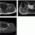
FIGURE 8.16 Osseous type choanal atresia in a newborn boy who presented with respiratory distress and inability to pass a nasogastric tube. Axial bone window CT image of the facial bones shows bony obliteration of both choanae with diamond-shaped vomers (arrows). Accumulated secretions (asterisks) are also present in the dependent potions of the nasal passages.


FIGURE 8.17 Membranous type of choanal atresia. Axial bone window CT image of the facial bones in a newborn girl who presented with respiratory distress shows narrowed posterior choanae on the right side with soft tissue density (arrow) compatible with right-sided membranous type of choanal atresia. Left-sided posterior choanae (asterisk) is normal and patent.

Active adenoid and tonsillar infection (with associated enlargement) is treated with antibiotics. For chronic
enlargement, particularly when associated with airway compromise or OSA, adenoidectomy and/or tonsillectomy may be required. Surgery may also help relieve associated recurrent sinus or ear infections.2


FIGURE 8.18 Congenital pyriform aperture stenosis in a newborn girl who presented with dyspnea while feeding. Axial bone window CT image of the facial bone shows marked narrowing (circled area) of the nasal passages anteriorly due to closed apposition of the maxilla anteriorly.


FIGURE 8.19 Adenoid, palatine, and lingual tonsillar hypertrophy in a 12-year-old girl who presented with progressively worsening oral breathing and snoring. Lateral radiograph of the upper airway shows moderate prominence of the adenoids (asterisk) causing narrowing of the nasopharynx. There is also substantial enlargement of the lingual tonsils (arrowhead) and palatine tonsils (arrow).


Macroglossia

Macroglossia refers to a chronic and painless enlargement of the tongue, which protrudes beyond the teeth or alveolar ridge at rest. It should be differentiated from acute glossitis, characterized by painful and rapid tongue enlargement.2,43 Causes include hypothyroidism, idiopathic hyperplasia, and various syndromes including the mucopolysaccharidoses, Down, and Beckwith-Wiedeman.2,44,45 A variant termed relative macroglossia refers to a proportionately large tongue in relation to a small mandible (micrognathia). Presenting symptoms include noisy breathing, drooling, dysphagia, and speech difficulty. Most concerning is the potential for upper airway obstruction.2,45,46

Sleep fluoroscopy can be used to assess progressive respiratory distress in affected pediatric patients. The enlarged tongue may be seen falling posteriorly during sleep and obstructing the posterior pharynx.2,41 Crosssectional imaging with CT and MRI is also useful in assessing the tongue and helping to exclude underlying masses2,47 (Fig. 8.20).


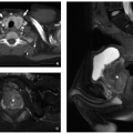
FIGURE 8.20 Macroglossia in a 4-year-old boy with Down syndrome and respiratory distress. A sagittal T2-weighted MR image shows a large tongue (asterisk) without abnormal internal signal. Glossoptosis or posterior displacement of the tongue is also seen (arrows).

In macroglossia related to an underlying systemic process, medical management may be sufficient. Otherwise, surgery with reduction glossectomy is the treatment of choice in symptomatic patients, helping to prevent future airway, speech, and orthodontic problems. Acute upper airway obstruction requires immediate attention and intervention such as tracheostomy.2,47


Laryngomalacia

Laryngomalacia is a benign, often transient, condition characterized by abnormal laxity of the pharyngeal soft tissues due to immaturity of the laryngeal cartilages and muscles. These abnormalities cause inspiratory collapse of the epiglottis, arytenoids, and aryepiglottic folds resulting in partial upper airway obstruction. Laryngomalacia is the most common congenital laryngeal anomaly. It is also the most common cause of symptomatic airway obstruction in infants presenting with stridor, which worsens with rest and improves with activity.2,48,49,50

Airway fluoroscopy characteristically demonstrates downward and posterior bending of the epiglottis and anterior buckling of the aryepiglottic folds, narrowing and eventually occluding the upper airway (Fig. 8.21). However, airway fluoroscopy, while relatively specific for the diagnosis, has poor sensitivity. Thus, even when normal, further evaluation with laryngoscopy should be performed if there is persistent clinical suspicion.2,48,51,52

Most cases of laryngomalacia are self-limited and resolve without intervention by the first year of life.2 Acid-suppressing medication should be given in patients with concurrent laryngomalacia and feeding symptoms.53 Persisting symptoms require surgical intervention to prevent complications such as airway obstruction and even sudden death. Current management techniques include supraglottoplasty, aryepiglottic fold incision, epiglottopexy, and tracheostomy.2,54


FIGURE 8.21 Laryngomalacia in a 2-month-old infant with stridor. A: Lateral radiograph of the upper airway demonstrates the normal position of the epiglottis (arrow). B: Lateral radiograph of the upper airway shows laxity of the epiglottis (arrow) with posterior and downward movement obstructing the airway. (Reprinted from Laya BF, Lee EY. Congenital causes of upper airway obstruction in pediatric patients: updated imaging techniques and review of imaging findings. Sem Roentgenol. 2012;47(2):147-158, with permission. Case courtesy of Khristine Grace C. Pulido, MD, Manila, Philippines.)


Tracheal Agenesis

Congenital tracheal agenesis is a rare and generally fatal anomaly. Although the pathogenesis is uncertain, it is postulated to result from failed embryologic separation of the trachea and esophagus during anterior budding of the proximal foregut.55 The estimated incidence is 1 in 50,000 newborns with a male/female ratio of 2:1.56 Over 150 cases have been reported since its initial description by Payne in 1900.56,57 More than 50% of affected patients are premature, and over half of pregnancies are associated with polyhydramnios.56,58 Associated congenital anomalies are present in 50% to 94% of cases. Affected pediatric patients typically present emergently with cyanosis, severe respiratory distress, inadequate gas exchange, lack of audible crying, and failed endotracheal intubation.56

Anatomically, the trachea is typically blind-ending below the level of the larynx, with gas exchange occurring through a distal esophageal fistula.59 As originally described by Floyd et al.,60 there are three recognized subtypes (Fig. 8.22). In type I, the proximal trachea is atretic. The preserved short distal trachea communicates with the esophagus via a tracheoesophageal fistula (TEF). Type II is most common, occurring in 60% of cases. The trachea is nearly or entirely absent. Two main bronchi join to form a midline carina, which most often fistulizes with the esophagus. In type III, the trachea and carina are absent. The main bronchi arise directly from the distal esophagus at separate origins.55,56,59,60

Chest radiographs are generally nondiagnostic but may demonstrate a missing tracheal air column, an abnormally low position of the tracheal bifurcation, an abnormally posterior “endotracheal” tube/esophageal intubation, or gaseous distention of the distal esophagus, stomach, and proximal small bowel (Fig. 8.23). Historically, barium studies were performed to assess for bronchoesophageal fistulas but are now generally avoided due to potential airway compromise.55,59,61 CT with multiplanar reformations (MPRs) is the test of choice, which can show the entire length of atresia and precise location of esophageal fistula.55,59

Few treatment options currently exist. If diagnosed prenatally by fetal ultrasound and MRI, the ex utero intrapartum tracheotomy (EXIT) procedure may be successful if the distal trachea is patent. Surgical attempts to reconstruct the upper airway using the esophagus or synthetic material thus far have been ineffective in allowing long-term survival.62


Tracheal Bronchus

Historically, tracheal bronchus was strictly defined as a right upper lobe bronchus arising from the trachea (also known as “bronchus suis” or pig bronchus due to similar morphology in pigs). The entity now encompasses a variety of bronchial anomalies originating from the trachea or main bronchi directed toward the upper lobes. Based on bronchographic and bronchoscopic series, the prevalence of right and left tracheal bronchus is 0.1% to 0.2% and 0.3% to 1%, respectively.1,7,63,64 Although generally isolated and incidental, reported associations include Down syndrome, rib anomalies, TEF, VATER (vertebral defects, anal atresia, tracheoesophageal fistula, esophageal atresia, renal defects, radial dysplasia) syndrome, partial anomalous pulmonary venous return, congenital lobar emphysema, and cystic lung malformations.63,64,65 Affected infants and children are usually asymptomatic but may present with recurrent pneumonia, atelectasis, or air

trapping, persistent cough, stridor, acute respiratory distress, or hemoptysis.63,64 A classic history is persistent right upper lobe atelectasis following endotracheal intubation because of obstruction of the unsuspected aberrant bronchus by the endotracheal tube.25,26


FIGURE 8.22 Diagram of three types of congenital tracheal agenesis. In congenital tracheal agenesis type I, the proximal trachea is atretic. The preserved short distal trachea communicates with the esophagus via a tracheoesophageal fistula. In congenital tracheal agenesis type II, the trachea is nearly or entirely absent. Two main bronchi join to form a midline carina, which most often fistulizes with the esophagus. In congenital tracheal agenesis type III, the trachea and carina are absent. The main bronchi arise directly from the distal esophagus at separate origins.


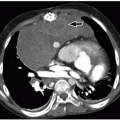
FIGURE 8.23 Tracheal agenesis in a 3-day-old boy with multiple congenital malformations including DiGeorge syndrome, tetralogy of Fallot, right aortic arch, and discontinuous pulmonary artery who presented with severe respiratory distress. A: Frontal chest radiograph shows a missing tracheal air column. B: Enhanced axial CT image shows an absent trachea and right mainstem bronchus (arrow) directly arising from the distended esophagus (E). Also noted is a nasogastric tube within the esophagus. (Case courtesy of Jonathan R. Dillman, MD, MSc, Cincinnati Children’s Hospital Medical Center, Cincinnati, OH.)

A “true tracheal bronchus” arises from the trachea generally <2 cm and no more than 6 cm from the carina, typically at the right lateral aspect.1,7,63,64 If the segmental bronchi of the parent anatomic bronchus supplying the same (upper) lobe as the tracheal bronchus are preserved, the tracheal bronchus is termed supernumerary. Otherwise, the tracheal bronchus is termed displaced. Blind-ending supernumerary bronchi are called tracheal diverticula. Supernumerary bronchi ending in aerated or bronchiectatic lung are called apical accessory lungs or tracheal lobes.64

Chest radiography may demonstrate the anomalous airfilled bronchus. However, CT is the preferred imaging modality for diagnosis. Newer postprocessing techniques such as 3D airway reconstruction and CT virtual bronchoscopy (CTVB) can help facilitate assessment1,66,67 (Fig. 8.24).

Asymptomatic patients require no treatment. However, surgical resection of the tracheal bronchus is recommended in those symptomatic.7 Additionally, preoperative recognition of tracheal bronchus helps optimize airway management; this is especially important in critically ill patients undergoing complex surgeries.66,68


FIGURE 8.24 Tracheal bronchus in an 11-month-old boy who presented with a history of recurrent right upper lobe atelectasis and infection. Subsequently obtained bronchoscopy confirmed an abnormal bronchus arising from the right lateral wall of the trachea. A: Enhanced axial soft tissue window CT image shows an anomalous right upper lobe bronchus (arrow), tracheal bronchus, arises directly from the lateral wall of the trachea (T). B: 3D external volume-rendered CT image of the large airways and lungs confirms the origin and course of the tracheal bronchus (arrow). C: 3D internal volume-rendered CT image shows an opening (arrow) of the tracheal bronchus located above the carina.


Tracheal Diverticulum

Tracheal diverticulum is a rare abnormality characterized by an outpouching of the posterolateral tracheal wall because of focal weakness of the trachealis muscle. The congenital form results from malformed supernumerary tracheal branches. The acquired form is caused by increased transluminal pressure within the trachea in patients with chronic cough or obstructive lung disease. Most affected pediatric patients are asymptomatic. However, large diverticula may harbor debris and cause recurrent pulmonary infections. Presenting symptoms also include foreign body sensation, neck or cervical swelling, and dysphagia.1,69,70,71,72,73 The Mounier-Kuhn syndrome (tracheobronchomegaly) is associated with multiple tracheal diverticula.74

CT with multiplanar 2D and 3D reformats accurately demonstrates a direct connection between the trachea and diverticulum, which can be difficult to visualize with bronchoscopy1,69,70,71,72,73 (Fig. 8.25). Diverticula more commonly occur on the right side; this may be related to the supporting esophagus and aortic arch on the left preventing diverticulum formation.72,73 Congenital diverticula are typically small and narrow mouthed, arising 4 to 5 cm below the true vocal cords, whereas acquired diverticula are larger and wide mouthed.1,69,70,71,72,73

Only gold members can continue reading. Log In or Register to continue

Stay updated, free articles. Join our Telegram channel

Oct 13, 2018 | Posted by in PEDIATRIC IMAGING | Comments Off on Airway

Full access? Get Clinical Tree

Get Clinical Tree app for offline access