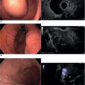
Christoph F. Dietrich Caritas‐Krankenhaus, Bad Mergentheim, Germany Radial and also longitudinal endorectal ultrasound (ERUS) allows precise imaging of the rectal wall in multiple planes and detailed information about the surrouding structures. The same is true for the generally not well known but effective anorectal ultrasound using a conventional transabdominal transducer technique (perineal ultrasound, PNUS). A transversal section with a 360‐degree transducer is most helpful for orientation and therefore for the examination of the anal canal, whereas for examination of more orally located structures (flexible or stiff) radial as well as sector scanners can be used. Sagittal, frontal and transverse, and other variable longitudinal sections have to be considered. The anatomical relationship between the internal anal sphincter (IAS), external anal sphincter (EAS), levator ani and puborectal muscles, and ischiorectal fossa can be reconstructed by the examiner’s anatomical knowledge and his or her aptitude for multidimensional imagination. Reproducible documentation and imaging can be also done using three‐dimensional (3D) techniques or real‐time 3D technique (also called 4D). A comprehensive categorization of the anal canal and distal rectum can be achieved by examining at different levels. From a didactic point of view, a step‐by‐step approach of retracting the transducer to indicate the distance in centimeters from the anocutaneous level seems to be helpful. A pushing forward technique may also be done but is less often used due to reduced reproducibility. Orientation is achieved by identification of the surrounding leading structures (bladder, prostate, vagina, puborectal muscle, etc.) and details are best seen with appropriate focusing and instrument adjustment (Video 8.1). Retracting the transducer, corresponding sections at 4–6 cm (perirectal tissue, prostate, seminal vesicles, cervix, vagina), at 2–4 cm (IAS, EAS, puborectal muscle), and at a superficial level of 0–2 cm (EAS with continuous fibers radiating into the subcutaneous fat) are visualized. Reporting of the anatomy and findings should always use clock orientation as a reference (i.e. the pubic symphysis is always at the 12 o’clock position). The anal canal is covered by a columnar epithelium below the anal crypts. The mucosa is a transitional cell epithelium and begins at the height of the anal crypts. It can, however, be only inconstantly visualized as a faintly dark layer with scattered echoes of medium echogenicity. The transitional epithelium disappears in the first echo‐rich border area depending on the type and frequency of the ultrasound probe. The dentate line and the anorectal crypts are of importance with respect to fistula formation but often cannot be seen mainly due to the compression of the anal canal by the endorectal probe. Normally, the hemorrhoidal plexus is virtually invisible, unless very thin probes are used or thrombosis is present. Important anatomical details are summarized in Table 8.1. The rectum can be examined at any level by endoscopic and/or visual orientation. The echogenicity of the various mural layers in the anal canal is similar to that in the gastrointestinal tract elsewhere and characterized by different, well‐separated bands when imaged from the lumen. Above the transition zone the typical sonographic features of the five (or nine) layered rectal wall are found. The typical five layers comprise the echo‐rich borderline between the probe and the mucosa, the echo‐poor mucosa, the echo‐rich submucosa, the echo‐poor muscularis propria, and the echo‐rich serosa and the border area to the perirectal fat tissue. Using highly sophisticated equipment up to nine layers can be displayed, including the two parts of the muscularis propria, differentiating the stratum circulare (inner ring layer) and the stratum longitudinale (outer ring layer) separated by a connective tissue‐like septum. A further differentiation of the inner layers yields additional layer separation in the mucosa and submucosa. In addition, it is relevant to note that these layer structures are important in staging rectal cancer according to the TNM classification. Table 8.1 Anatomical structures and abbreviations. The standardized approach starts at a level of 4–6 cm above the anocutaneous line (ab ano) by delineating the seminal vesicles, prostate, and urethra in men, whereas at this level the cervix uteri and vagina can be displayed in women, and the urinary bladder in both. The internal iliac vessels cross the rectosigmoid colon just proximal to this region, but definite determination of the edge between the rectum and sigmoid colon is endosonographically still an unsolved problem. The surrounding bones and muscles of the pelvic floor, namely the levator ani muscles (ischiococcygeus, iliococcygeus, and pubococcygeus) including the V‐shaped puborectal muscle (Figure 8.1), should be identified since the anal canal ends at this level and the rectum begins. The funnel‐like structure of the levator ani muscle separates the extraperitoneal pelvis into an infraperitoneal or pelvirectal and a subcutaneous or ischiorectal space. The lateral border of the pelvic floor corresponds to the M. obturatorius internus with the foramen obturatorium. The second section at 2–4 cm ab ano shows as a leading structure the prominent EAS as a 5–10 mm broad layer characterized by often concentrically arranged echo‐rich and sometimes inhomogeneous series of lines (Video 8.1 and Figure 8.2) depending on the examination technique used (criteria: radial or linear probes, diameter of the probe, transducer frequency, tissue harmonic imaging, focus zone). The EAS is composed of three more or less separated parts, i.e. the subcutaneous (pars subcutanea), the superficial (pars superficialis), and the deep (pars profunda) components which are confluent with each other. It is of great importance to understand that the shape and configuration of the EAS differs significantly between males and females. The EAS continues into the puborectal muscle (PRM) at the cranial margin of the anal canal, which is of importance for the delineation of ischiorectal and pelvirectal fossae. More orally the muscular structures are no longer vertically imaged so that this part of the muscle yields a weaker and less defined image (Video 8.1). At this level the os coccygeum with the coccygeal ligament can be also delineated. Ventrally this broad muscle layer reaches the inferior part of the pubic bone with inclusion of the urethra and the inferior part of the prostate or the vagina, respectively. Figure 8.1 (a) Levator ani muscle with the puborectal part (PR) in between markers. (b) The more distant parts of this muscle often show a more inhomogeneous and hypoechoic appearance (Hypo). Figure 8.2 Internal anal sphincter (echo‐poor) and external anal sphincter (echo‐rich). The superficial layer of the anal canal is also indicated (MUC). Coming back to gender differences, one has to keep in mind that the anal canal is generally 0.5–1.0 cm longer in males and the three different parts of the EAS are configured cylindrically around the anus, which means that the muscle shape and length is roughly the same ventrally and dorsally. In contrast, the three sections of the EAS are fused to a narrow band ventrally at the level of the pars superficialis in females. The notion of this anatomical difference is of importance in order not to misinterpret normal anatomical findings. Normal sphincter values are highly dependent on age, sex, and examination technique parameters (Table 8.2). The less pronounced echo‐poor IAS can be visualized more distally than the PRM and EAS (see below). The IAS can regularly be seen as a homogeneous echo‐poor layer with a thickness of 2–4 mm, without any relevant gender‐dependent differences, a little more distally than the EAS (Figure 8.2). The anal mucosa and the IAS muscle are considered the continuation of the inner rectal ring muscle. However, the measured thickness depends on the probes used, as an increasing probe diameter will lead to a thinning of the surrounding structures. The echo texture and thickness of the IAS tend to increase with age. The normal thickness, however, does not exceed 4 mm even in the elderly. Table 8.2 Normal sphincter values. This is highly dependent on age, sex, and probe diameter, since the measured thickness depends on the probes used, as an increasing probe diameter will lead to a thinning of the surrounding structures. The intersphincteric space with fibers of the corrugator (or longitudinal) muscle as a continuation of the rectal external longitudinal muscle layer is seen endosonographically as a thin echo‐poor layer containing echo‐rich connective tissue septa representing the border areas of the echo‐poor IAS and the first less echogenic layer of the EAS. It can be identified a little above 2 cm from the anocutaneous line. At this level the fossa ischiorectalis, the anococcygeal ligament, and the os coccygeum can be visualized. Additional points of orientation at this level are the radix penis with the ischiocavernosus muscle and the ramus ossis ischii. At this level also the echo‐rich border area between the ultrasound probe and the mucosa (or the anal cutis), the echo‐poor extremely thin mucosa which often cannot be recognized as a defined layer due to a lacking muscularis and its very thin diameter, and the echo‐rich submucosa (and subcutis) can be differentiated leading to the structures of the lowest level. The last section shows the cutis, the transitional epithelium, and the subcutis with the transition to the submucosa and more orally the IAS surrounded by the superficial and profound transverse perineal muscle and the fossa ischiorectalis as already described. The submucosa and the subcutaneous connective tissue appear as a homogeneous echogenic layer increasing in thickness from the superior to the inferior margin of the anal canal. The transversus perinei profundus and superficialis muscles and the ischiocavernosus muscle can also be imaged by ERUS.
8
Anatomy of the Anorectum: Radial and Linear
Introduction
Examination technique
Orientation
Normal anatomy
Anatomical remarks
Rectal wall
ARUS
Anorectal endosonography (alternative: perineal ultrasound, PNUS)
ERUS
Endorectal endosonography
C
Cutis (skin)
Muc
Mucosa
IAS
Internal anal sphincter. Musculus sphincter ani internus (inner [ring] muscle fibers of the rectum radiating into M. sphincter ani internus)
EAS
External anal sphincter. M. sphincter ani externus
LAM
Levator ani muscle (M. puborectalis, M. ischiococcygeus, M. iliococcygeus, M. pubococcygeus; endosonographic separation of the muscle layer is not always possible)
LM
Longitudinal muscle, M. corrugator ani (longitudinal muscle fibers of the rectum radiating into M. sphincter ani externus)
Level of the prostate or cervix uteri
External anal sphincter level


Internal anal sphincter level
M. sphincter ani internus
1.5–4.0 mm
M. sphincter ani externus
5–10 mm
Cutis and subcutis
Stay updated, free articles. Join our Telegram channel

Full access? Get Clinical Tree


