Intracranial anatomy is highly complex and well beyond the scope of this book. However, a number of structures are closely related to visual function and form the neural pathways for visual signals from the retina to the striate cortex. Associated neural and bony structures adjacent to this pathway are the sites for many pathologic processes that can affect the visual system. Here, we will only briefly review these visual pathways and the most important associated structures as a basis for the radiologic interpretation of diseases affecting visual function.
The skull base
The skull base forms the floor of the cranial cavity, and serves as a major interface between the intracranial compartment and the facial structures. Five bones contribute to the skull base, three unpaired midlines bones – ethmoid, sphenoid, and occipital bones – and the paired temporal bones ( Figure 4.1 ). While the frontal bone is generally unpaired in the adult, embryologically it begins as a paired structure and in young children the remains of the frontal or metopic suture may sometimes be seen between the brow ridges. The frontal bone forms the floor of the anterior cranial fossa.

The skull base begins immediately behind the frontal sinus and extends posteriorly to the clivus, formed by the posterior sphenoid and the basioccipital bones just in front of the foramen magnum. A number of structures of radiologic interest along the base serve as landmarks for local anatomy. Just posterior to the cribriform plate is the planum sphenoidale and anterior clinoid processes. Most of the floor of the anterior cranial fossa is formed by the orbital portion of the frontal bone on either side of the midline. It is grooved, reflecting the overlying gyri of the frontal lobe. Lying in the midline is the cribriform plate of the ethmoid bone, a perforated lamina of bone transmitting fibers of the olfactory nerve. On coronal sections, the cribriform plate is the lowest part of the anterior fossa, and may be as much as 1 cm lower than the roof of the ethmoid sinus. A thin vertical plate of bone, the crista galli, extends upward from the cribriform plate in the midline and serves for attachment of the falx cerebri between the cerebral hemispheres.
The chiasmatic sulcus is a small depression between the clinoid processes and houses the optic chiasm. The anterior wall of the sulcus is formed by the body of the sphenoid, and the lateral walls by the lesser wings. The anterior clinoids form the posterolateral borders. These bony structures are identifiable landmarks for the chiasm and the supracavernous portion of the internal carotid artery (ICA).
Sella turcica
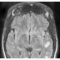
The body of the sphenoid bone makes up the central portion of the middle cranial fossa and skull base, and it contains the sella turcica. The sella, or pituitary fossa, lies between the anterior and posterior clinoid processes and is a depression that houses the pituitary gland ( Figure 4.2 ). The tuberculum sellae is a small elevation between the clinoids and just behind the chiasmatic sulcus; the pituitary fossa is the deepest portion and the posterior part of the sella is the dorsum sellae.

Cranial foramina
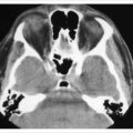
A number of foramina provide access into or out of the cranial cavity for neural, vascular, and other elements. These are useful to recognize as they provide important anatomic landmarks. Four openings lie along a line from the anterior clinoids posterolaterally toward the petrous pyramid. These are the superior orbital fissure, foramen rotundum, foramen ovale, and the foramen spinosum ( Figures 4.1–4.3 ).

The superior orbital fissure (SOF) begins just lateral to the anterior clinoid process and extends inferomedially between the greater and lesser sphenoid wings. On axial orbital scans it appears as an opening between the orbit and the middle cranial fossa. The SOF transmits all of the motor and sensory nerves of the orbit, with the exception of the optic nerve. As discussed in Chapter 3 , the central portion of the fissure is bridged by the annulus of Zinn, dividing it into three portions. The trochlear nerve (cranial nerve IV), the superior ophthalmic vein, and the frontal and lacrimal branches of the superior division of the trigeminal nerve (V 1 ) pass through the SOF above the annulus. The oculomotor nerve (cranial nerve III), abducens nerve (cranial nerve VI), and the nasociliary branch of V 1 enter the orbit through the annulus. The inferior ophthalmic vein branches pass to the cavernous sinus through the SOF below the annulus of Zinn.
The foramen rotundum is located in the sphenoid just below the SOF just lateral to the sella turcica. It transmits the maxillary division of the trigeminal nerve (cranial nerve V 2 ) into the pterygopalatine fossa, which is situated just below the inferior orbital fissure. About 1 cm posterolateral to the foramen rotundum is the foramen ovale in the base of the greater sphenoid wing, just in front of the petrous portion of the temporal bone. This carries the mandibular division of the trigeminal (cranial nerve V 3 ), the accessory meningeal artery, the lesser superficial petrosal nerve, and collateral veins to the pterygoid venous plexus in the infratemporal fossa. The foramen spinosum lies immediately posterior to the foramen ovale and transmits the middle meningeal artery from the external carotid system to supply the dura.
At the junction of the petrous bone and the occipital bone is the large jugular foramen. It transmits the glossopharyngeal (cranial nerve IX), vagus (cranial nerve X), and accessory (cranial nerve XI) nerves. It also transmits the inferior petrosal and transverse sinuses en route to the internal jugular vein. Externally its anterior portion is the carotid canal, but the ICA and internal jugular vein diverge as they enter the cranial cavity.
On the posterior surface of the petrous bone, above the jugular foramen is the acoustic foramen transmitting the facial nerve (cranial nerve VII), acoustic nerve (cranial nerve VIII), and the nervus intermedius branch of the facial nerve, which carries the special sensory fibers for taste. Just inferomedial to the jugular foramen is the small hypoglossal foramen which transmits the hypoglossal nerve (cranial nerve XII).
Cavernous sinus
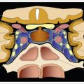
The cavernous sinus is a venous plexus within a split in the dura on either side of the body of the sphenoid bone. Anatomically the cavernous sinus begins at the cranial opening of the superior orbital fissure and extends posteriorly to the petrous portion of the temporal bone. Within the dura of the sinus walls run several cranial nerves subserving visual function ( Figure 4.4 ). Along the medial wall runs the ICA and just lateral to it is the abducens nerve (cranial nerve VI). Within the lateral sinus wall from superior to inferior are the oculomotor nerve (cranial nerve III), trochlear nerve (cranial nerve IV), and the ophthalmic (cranial nerve V 1 ) and maxillary divisions (cranial nerve V 2 ) of the trigeminal nerve. In the anterior-most part of the cavernous sinus the oculomotor nerve divides into its superior and inferior divisions before entering the superior orbital fissure en route to the orbital apex.