Chapter contents
- I.
COMPUTED TOMOGRAPHY SCANS 50
- A.
Computed tomography: axial cranial bone anatomy 50
- B.
Computed tomography: coronal cranial bone anatomy 56
- C.
Computed tomography: sagittal cranial bone anatomy 62
- D.
Computed tomography: axial intracranial soft tissue anatomy 67
- E.
Computed tomography: axial orbital soft tissue anatomy 72
- F.
Computed tomography: coronal orbital soft tissue anatomy 76
- G.
Computed tomography: sagittal orbital soft tissue anatomy 81
- A.
- II.
MAGNETIC RESONANCE IMAGING SCANS 86
- A.
Magnetic resonance imaging: axial intracranial soft tissue anatomy 86
- B.
Magnetic resonance imaging: coronal intracranial soft tissue anatomy 91
- C.
Magnetic resonance imaging: axial orbital soft tissue anatomy 97
- D.
Magnetic resonance imaging: coronal orbital soft tissue anatomy 102
- E.
Magnetic resonance imaging: sagittal orbital soft tissue anatomy 106
- F.
Magnetic resonance imaging: the suprasellar cistern 110
- G.
Magnetic resonance imaging: MR angiography 114
- H.
Magnetic resonance imaging: MR venography 116
- I.
Magnetic resonance imaging: the cavernous sinus 119
- A.
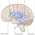
In the following pages a series of computed tomography (CT) and magnetic resonance (MR) images are presented at various orientations with the most important anatomic features labeled. The reader can refer back to Chapters 3 and 4 for anatomic reference.
I
Computed tomography scans
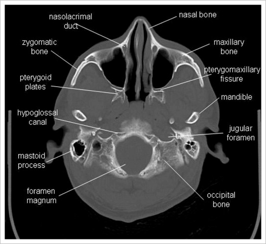
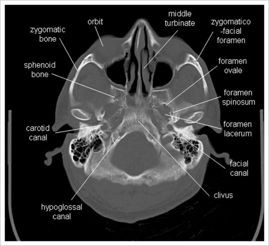
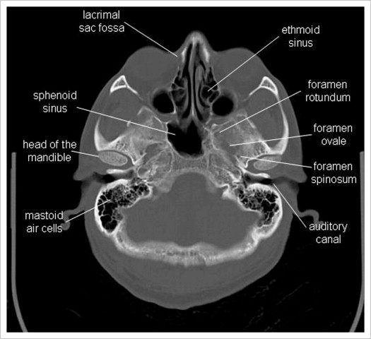
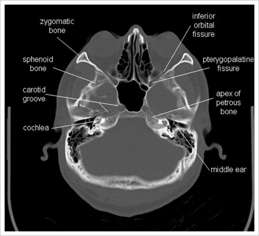
A
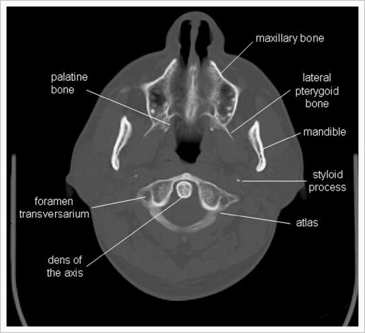
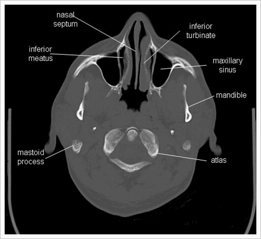
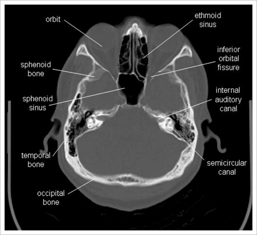
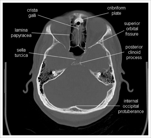
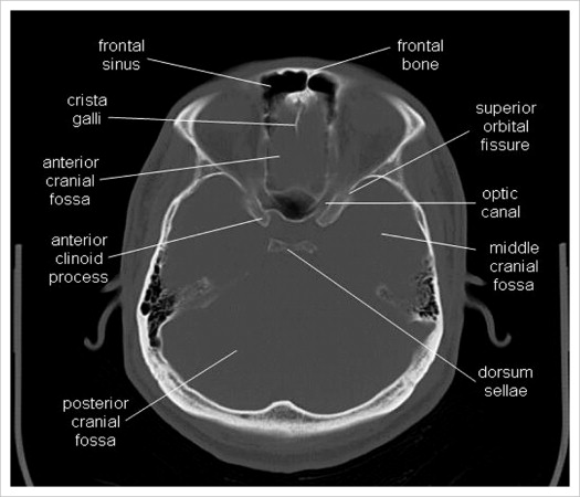
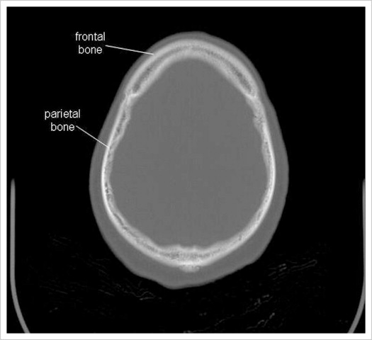
Computed tomography: axial cranial bone anatomy

B
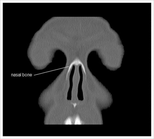
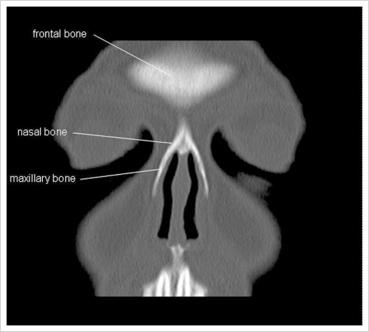
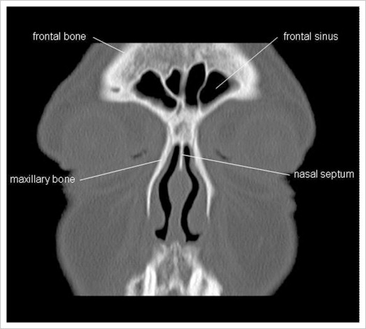
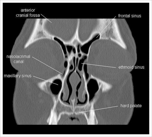
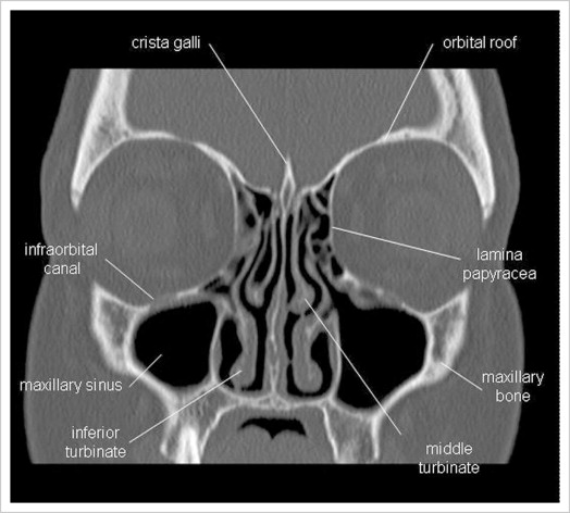
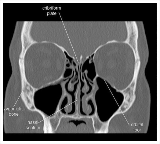
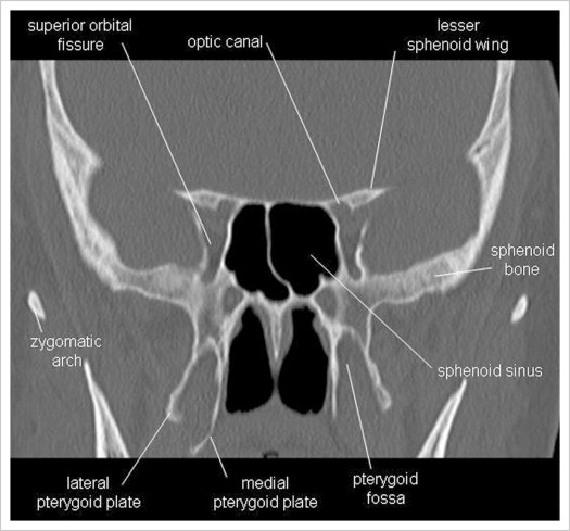
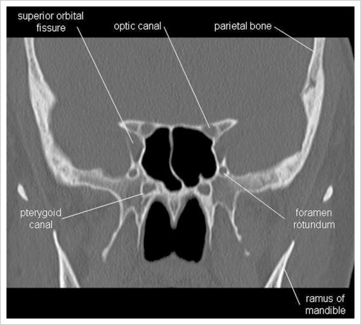
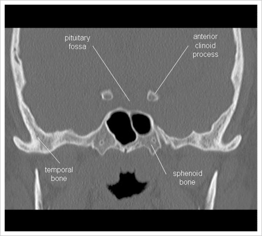
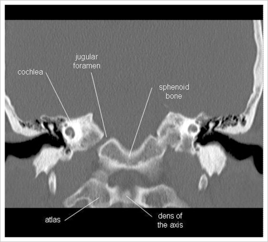
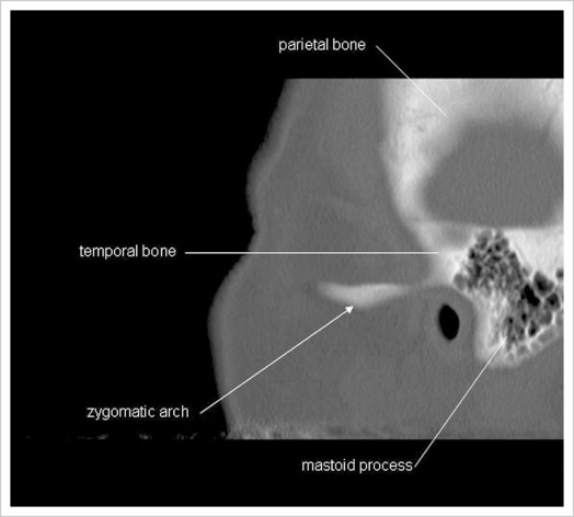
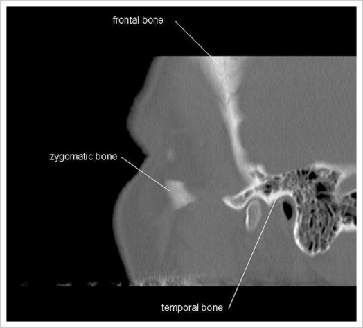
Computed tomography: coronal cranial bone anatomy


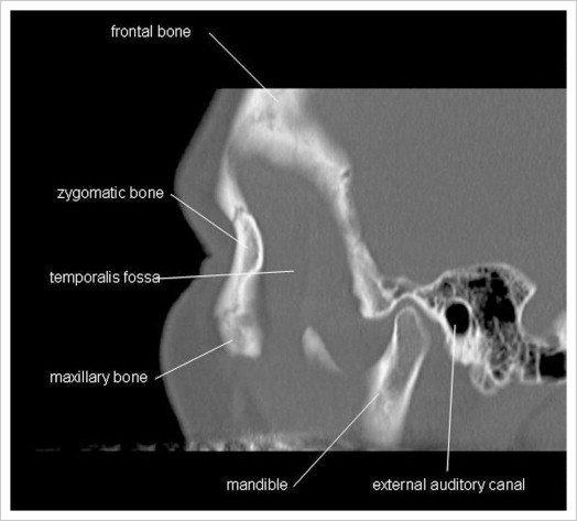
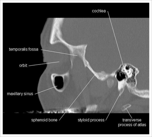
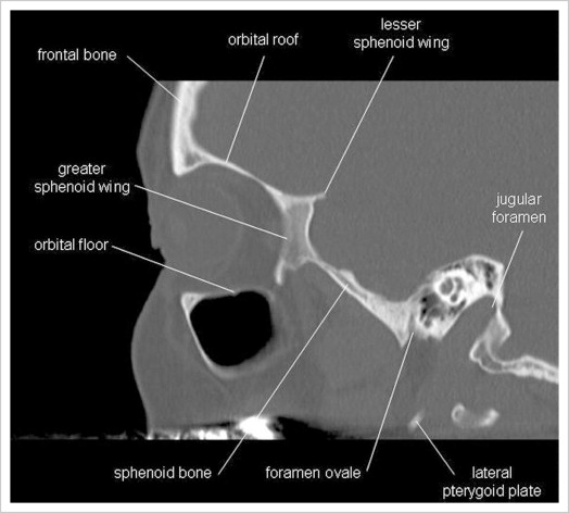
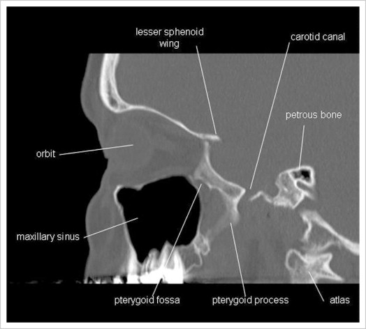
C
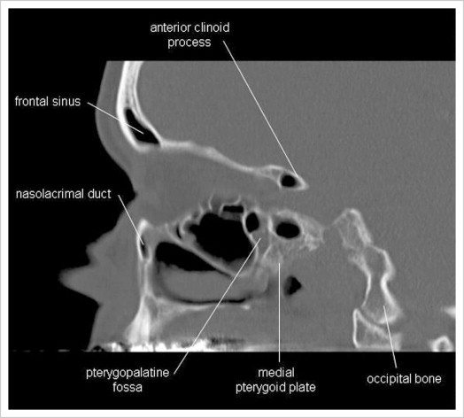
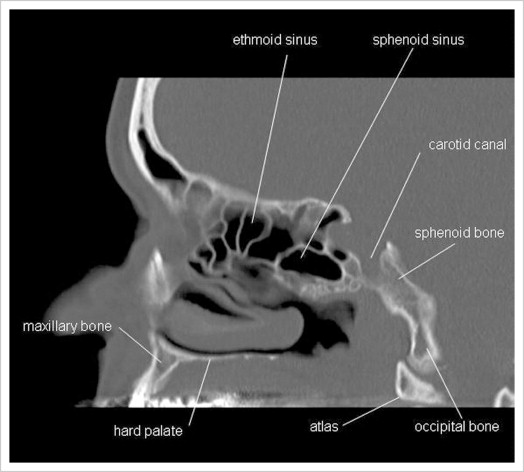
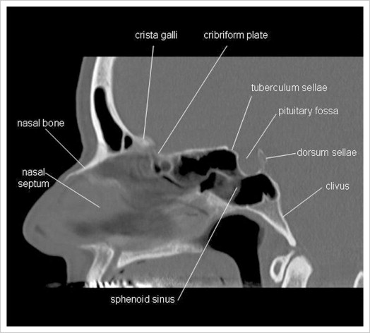
Computed tomography: sagittal cranial bone anatomy
D
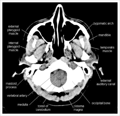
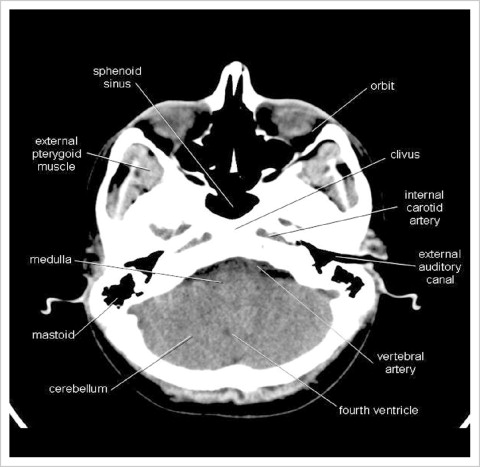
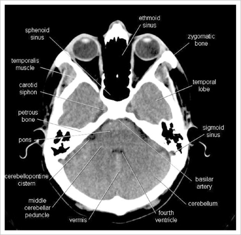
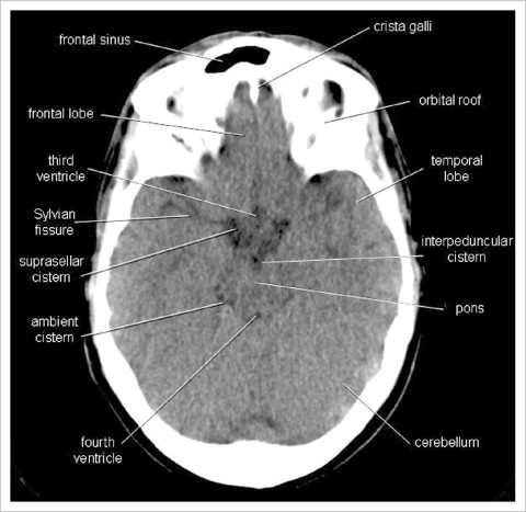
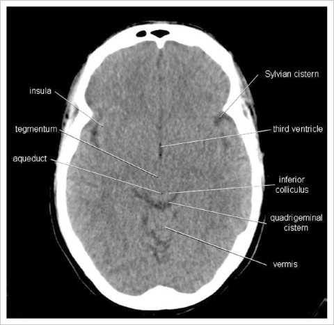
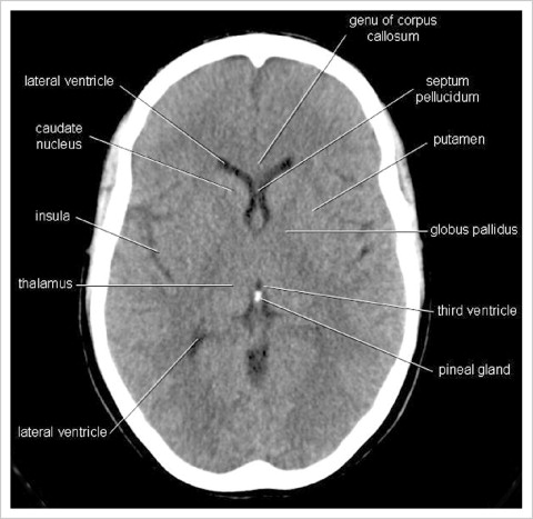
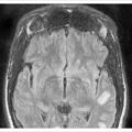
Computed tomography: axial intracranial soft tissue anatomy