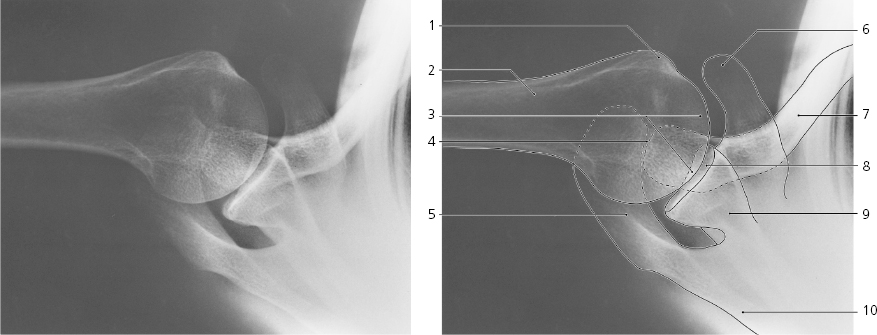
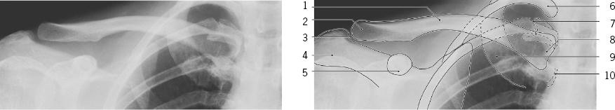
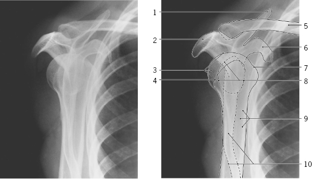
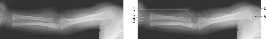
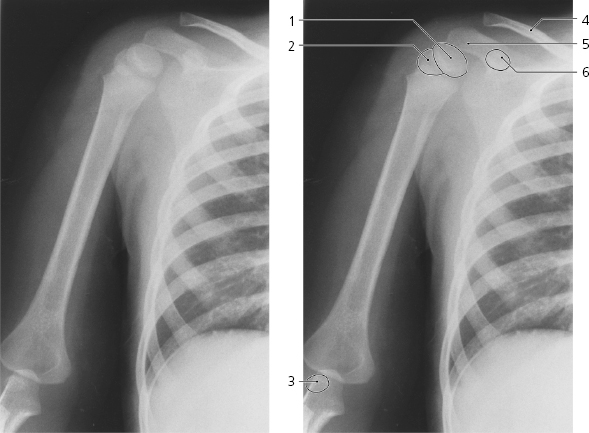
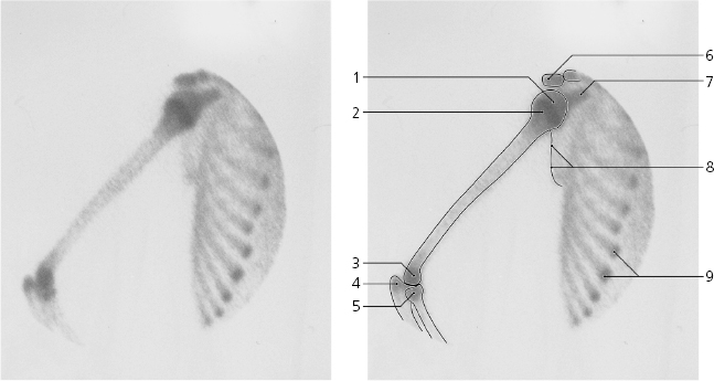
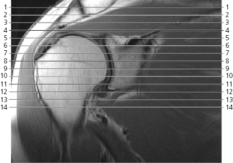
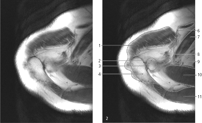
Shoulder, a-p X-rayAcromial end of clavicle Acromion Humeral head Epiphyseal scar Anatomical neck Greater tubercle Lesser tubercle Coracoid process Surgical neck Glenoid cavity Neck of scapula Lateral border of scapula Spine of scapula First rib Superior angle of scapula Medial border of scapula Sternal end of clavicle Manubrium of sternum Sternal angle Body of sternum Inferior angle of scapula Shoulder , axial X-rayGreater tubercle Surgical neck of humerus Humeral head Acromioclavicular joint Acromion Coracoid process Clavicle Glenoid cavity Neck of scapula Spine of scapula Clavicle, a-p X-rayShaft of clavicle Acromial end of clavicle Acromioclavicular joint Acromion Coracoid process Second rib Costotransverse joint Sternal end of clavicle First rib Costovertebral joint Scapula, oblique X-raySuperior margin of scapula Acromion Head of humerus Greater tubercle Clavicle Coracoid process Lesser tubercle Glenoid cavity Surgical neck of humerus Scapula from edge Shoulder and arm, a-p X-ray, child one yearCapitulum (ossification center) Capitate bone (ossification center) Hamate bone (ossification center) Greater tubercle (ossification center) Humeral head (ossification center) Shoulder and arm , a-p X-ray, child 5 yearsHumeral head (ossification center) Greater tubercle (ossification center) Capitulum (ossification center) Clavicle Acromion Coracoid process (ossification center) Shoulder and arm , 99m Tc-MDP, scintigraphy, child 12 yearsHumeral head Growth plate of proximal epiphysis of humerus Trochlea and capitulum Olecranon Head of radius Acromion Coracoid process Lateral margin of scapula Osteochondral transition of ribs Scout view of shoulder Shoulder , axial MRDeltoideus → Acromioclavicular joint with articular disc → Acromion → Clavicle → Coracoclavicular (trapezoid) ligament (attachment) → Suprascapular artery and vein → Supraspinatus → Trapezius → Shoulder , axial MR
Scout view on page 52Deltoideus ↔ Clavicle (acromial extremity) ← Acromioclavicular joint ← Acromion ↔ Spine of scapula → Thoracoacromial artery/vein Subclavius muscle Coracoclavicular (trapezoid) ligament ↔ Coracoclavicular (conoid) ligament → Supraspinatus ↔ Trapezius ↔ Pectoralis major → Cephalic vein → Suprascapular artery/vein ← Shoulder , axial MR
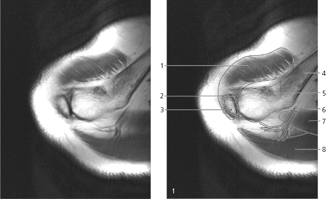
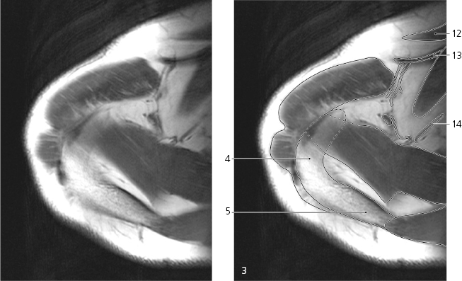
Scout view on page 52Coracoacromial ligament → Acromion ← Spine of scapula ↔ Greater tubercle of humerus → Head of humerus → Deltoideus ↔ Infraspinatus → Pectoralis major ↔ Cephalic vein ↔ Clavipectoral fascia Trapezoid and conoid ligament (attachment on coracoid process) ← Supraspinatus ↔ Trapezius ↔ Pectoralis minor → Coracoacromial ligament (attachment) ← Coracoid process → Articular capsule/rotator cuff →
Only gold members can continue reading.
Log In or
Register to continue
Stay updated, free articles. Join our Telegram channel
Join